. 2015 May 15;290(20):12487-96.
doi: 10.1074/jbc.M114.607580. Epub 2015 Mar 13.

Histone deacetylase 6 (HDAC6) promotes the pro-survival activity of 14-3-3ζ via deacetylation of lysines within the 14-3-3ζ binding pocket

Affiliations

Histone deacetylase 6 (HDAC6) promotes the pro-survival activity of 14-3-3ζ via deacetylation of lysines within the 14-3-3ζ binding pocket

Jeffrey B Mortenson et al. J Biol Chem. .

Abstract

The phospho-binding protein 14-3-3ζ acts as a signaling hub controlling a network of interacting partners and oncogenic pathways. We show here that lysines within the 14-3-3ζ binding pocket and protein-protein interface can be modified by acetylation. The positive charge on two of these lysines, Lys(49) and Lys(120), is critical for coordinating 14-3-3ζ-phosphoprotein interactions. Through screening, we identified HDAC6 as the Lys(49)/Lys(120) deacetylase. Inhibition of HDAC6 blocks 14-3-3ζ interactions with two well described interacting partners, Bad and AS160, which triggers their dephosphorylation at Ser(112) and Thr(642), respectively. Expression of an acetylation-refractory K49R/K120R mutant of 14-3-3ζ rescues both the HDAC6 inhibitor-induced loss of interaction and Ser(112)/Thr(642) phosphorylation. Furthermore, expression of the K49R/K120R mutant of 14-3-3ζ inhibits the cytotoxicity of HDAC6 inhibition. These data demonstrate a novel role for HDAC6 in controlling 14-3-3ζ binding activity.

Keywords: 14-3- Protein; Apoptosis; Cell Signaling; Histone Deacetylase 6 (HDAC6); Metabolic Stress; Non-histone Acetylation; Stress.


Figures

FIGURE 1.
FIGURE 1.
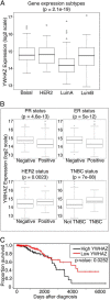
14-3-3ζ expression is elevated in receptor-negative breast tumors, and high expression correlates with decreased patient survival. A, YWHAZ (14-3-3ζ) expression was analyzed in basal (n = 94), HER2 (n = 56), LumA (n = 228), and LumB (n = 123) breast cancer samples and expressed as Box-and-Whisker plots. p value was calculated by comparing all groups with the analysis of variance test. B, 14-3-3ζ expression was analyzed in progesterone receptor (PR) positive (n = 523), PR negative (n = 252), estrogen receptor (ER) positive (n = 602), ER negative (n = 176), HER2 positive (n = 129), HER2 negative (n = 429), triple negative breast cancer (TNBC, n = 85), and non-TNBC (n = 468) data sets. Data are expressed in Box-and-Whisker plots. p values were calculated using standard two-sample t test. C, Kaplan Meier analysis of 14-3-3ζ expression levels correlated with patient survival. Breast cancer patients whose tumors fell within the highest (n = 164) and lowest (n = 163) quintiles of 14-3-3ζ expression were compared. Statistical significance was calculated using the log-rank test. All data were derived from The Cancer Genome Atlas (TCGA) database.
FIGURE 2.
FIGURE 2.
Acetylated lysines within the 14-3-3ζ binding pocket and protein-protein interface. A, three-dimensional x-ray crystal structure of 14-3-3ζ homodimer in gold (PDB code 4IHL) bound to a c-Raf phosphoserine motif (green). Inset is a magnification of 14-3-3ζ binding pocket. Positively charged Lys49 and Lys120 of 14-3-3ζ are closely associated with negatively charged phosphate group of c-Raf. The crystal structure was rendered using Chimera software (UCSF) from a published 14-3-3 structure at a resolution of 2.20 Å (32). Four acetylated lysine residues, identified by mass spectrometry, are labeled (K49, K120, K193, and K212). B, amino acid sequence alignment of regions surrounding Lys49 and Lys120 (human numbering) from the indicated organisms. Accession numbers: human, NP_663723; rat, NP_037143; zebrafish, NP_997922; potato, BAM28646. Of note, potato (Solanum tuberosum) 14-3-3 is not designated as the ζ isoform but rather a “14-3-3-like protein”.
FIGURE 3.
FIGURE 3.
Acetylation-mimicking mutations abolish 14-3-3ζ interactions. A, HEK-293T cells were transfected with WT or mutant forms of HA-tagged 14-3-3ζ plasmid expression vector (Lys to Gln mutants at each acetylated lysine residue). Cell lysates were immunoprecipitated with HA-agarose resin and run on 12% SDS-PAGE for immunoblotting for kinesin, AS160, Liprin-β (36), and Bad. Representative data are of at least three replicate experiments. B, HEK-293T cells were transfected with WT or K49Q HA-tagged 14-3-3ζ and subjected to gel filtration. Eluted fractions were collected and run on separate 12% SDS-PAGE for immunoblotting with HA (14-3-3ζ) antibody. Representative data are of at least four replicate experiments.
FIGURE 4.
FIGURE 4.
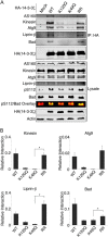
Deacetylation-mimicking mutations rescue the loss of 14-3-3ζ interactions. A, HEK-293T cells were transfected with WT or mutant forms of HA-tagged 14-3-3ζ plasmid expression vector (Lys to Gln mutants at Lys49 or Lys120, Lys to Arg double mutant at Lys49 and Lys120). Cell lysates were immunoprecipitated with HA-agarose resin and run on 12% SDS-PAGE for immunoblotting of the binding partners kinesin, AS160, Liprin-β, Bad, and Atg9 (36). Phosphorylation of the 14-3-3ζ binding site of Bad (Ser112) was compared with total Bad in lysate (overlay). Representative data from seven replicate experiments. B, Western blot data from panel A were quantified using LI-COR infrared band imaging. Relative interaction values represent the quantified signal of interacting-protein divided by HA signal. p values indicated by asterisk were calculated using two-sample t test and are as follows: kinesin = 0.042; Liprin-β = 0.044; Bad = 0.041.
FIGURE 5.
FIGURE 5.
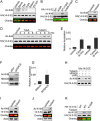
HDAC6 deacetylates 14-3-3ζ at Lys49 and Lys120. A, HEK-293T cells were transfected with WT HA-tagged 14-3-3ζ expression vector and treated with various HDAC inhibitor drugs (SAHA, 10 μm; trichostatin A (TSA), 10 μm; tubastatin A (TubA), 40 μm; salermide, 40 μm; and EX-527, 40 μm) or vehicle (dimethyl sulfoxide) under normal conditions for 9 h. Cell lysates were immunoprecipitated with HA resin and run on 12% SDS-PAGE immunoblotting for Ac-K49 (green) and HA (14-3-3ζ, red). Representative data are of at least three replicate experiments. B, HEK-293T cells overexpressing WT or K49R mutant HA-tagged 14-3-3ζ were treated with tubastatin A (40 μm) or vehicle for 9 h followed by HA immunoprecipitation and immunoblot as described in the legend to Fig. 4A (exception: HA is in the green channel, Ac-K49 is in the red channel). Representative data are of at least four replicate experiments. C, U2OS cells overexpressing WT HA-tagged 14-3-3ζ were treated with tubacin (30 μm) for 8 h followed by HA immunoprecipitation and immunoblot as described in the legend to Fig. 4A. Representative data are of at least three replicate experiments. D, HEK-293T cells overexpressing WT HA-tagged 14-3-3ζ were treated with tubacin (35 μm) or tubastatin A (35 μm) in a 2-, 4-, 6-, and 8-h time course followed by HA immunoprecipitation and immunoblot as described in the legend to Fig. 4A. Representative data are of at least three replicate experiments. E, 8 h HDAC6 inhibitor time points from four replicate experiments of panel D were quantified. Relative acetylation signal is acetyl-Lys49 signal divided by HA signal. Bands were quantified by LI-COR infrared imaging. F, HEK-293T cells overexpressing WT HA-14-3-3ζ were transfected with HDAC6 siRNA (75 nm) or scrambled siRNA (75 nm). Cell lysates were HA immunoprecipitated and immunoblotted as in A. Representative data are of at least three replicate experiments. G, three replicate experiments from panel F were quantified as in panel E. H, recombinant His-tagged 14-3-3ζ was expressed in BL21-DE3 Escherichia coli and purified on a nickel resin. Resin-bound 14-3-3ζ was incubated with p300 acetyltransferase followed by vehicle, HDAC6 recombinant enzyme, or HDAC6 enzyme and tubacin (40 μm). 14-3-3ζ was analyzed by 12% SDS-PAGE and immunoblot with Ac-Lys49 and His (14-3-3ζ). Representative data are of at least three replicate experiments. I, MDA-MB-231 breast cancer cells were treated with tubacin (30 μm) or vehicle for 8 h. Endogenous 14-3-3ζ was analyzed by resolving cell lysate (40 μg) using 12% SDS-PAGE and immunoblotting with Ac-Lys120 (green), 14-3-3ζ (red), and actin. Representative data are of at least three replicate experiments. J, MDA-MB-231 breast cancer cells were treated as described in the legend to Fig. 4G. Endogenous 14-3-3ζ was analyzed as described in the legend to Fig. 4G. Representative data are of at least three replicate experiments. K, HEK-293T cells overexpressing WT or K120R mutant HA-tagged 14-3-3ζ were treated with tubacin (40 μm) or vehicle for 9 h followed by HA immunoprecipitation and immunoblot with Ac-Lys120 (green) and HA (red). Representative data are of at least three replicate experiments.
FIGURE 6.
FIGURE 6.
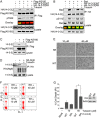
Inhibition of HDAC6 triggers a loss of 14-3-3ζ interactions and an increase in cell death, both of which are rescued by the K49R/K120R double mutant. A, HEK-293T cells were co-transfected with WT FLAG-tagged AS160 expression plasmid along with either WT HA-tagged 14-3-3ζ, or K49R/K120R (RR) double mutant expression plasmid, followed by treatment with tubacin (30 μm) or vehicle for 6 h. Cell lysates were immunoprecipitated using FLAG-agarose and run on 12% SDS-PAGE for immunoblot with HA (14-3-3ζ), phosphothreonine 642 (pT642), and FLAG (AS160). Representative data are of at least three replicate experiments. B, HEK-293T cells were co-transfected with Bad expression plasmid along with WT HA-tagged 14-3-3ζ, or K49R/K120R (RR) double mutant expression plasmid, followed by treatment with tubacin (30 μm) or vehicle for 9 h. Cell lysates were immunoprecipitated using HA-agarose and run on 12% SDS-PAGE for immunoblot with Bad and HA (14-3-3ζ). Cell lysate was immunoblotted with phosphoserine 112 (pS112). Representative data are of at least three replicate experiments. C, HEK-293T cells were transfected with WT FLAG-tagged AS160 expression plasmid or mock followed by treatment with tubacin (40 μm) or vehicle for 6 h. Cell lysates were immunoprecipitated using FLAG-agarose and run on 12% SDS-PAGE for immunoblot with 14-3-3ζ and FLAG (AS160). Representative data are of at least four replicate experiments. D, HEK-293T cells were transfected with WT HA-tagged Atg9 expression plasmid or mock followed by treatment with tubacin (40 μm) or vehicle for 6 h. Cell lysates were immunoprecipitated using HA-agarose and run on 12% SDS-PAGE for immunoblot with 14-3-3ζ and HA (Atg9). Representative data are of at least four replicate experiments. (E) HEK-293T cells overexpressing either WT 14-3-3ζ, K49R/K120R (RR) double mutant 14-3-3ζ, or mock transfection were treated with tubacin (10 μm, 40 μm) or vehicle for 48 h. Cell death was measured by PI staining and flow cytometry. Data are presented as light microscope pictures (scale bar = 250 μm); F, flow cytometry dot plots; G, bar graph (mean ± S.E., n = 3, p value = 0.029). p value was calculated using two-sample t test. Inset is a Western blot comparing expression of WT or RR mutant 14-3-3ζ. Cell lysates were analyzed via 12% SDS-PAGE and Western blot. Membrane was probed for HA (14-3-3ζ) and actin.
FIGURE 7.
FIGURE 7.
HDAC6 maintains 14-3-3ζ (shown as a dimer) binding activity by deacetylating Lys49 and Lys120 within the 14-3-ζ binding pocket. Inhibition of HDAC6 leads to 14-3-3ζ acetylation and loss of 14-3-3ζ binding activity. Acetylation-induced dissociation of 14-3-3ζ interactions (e.g. with Bad and AS160) results in a loss of pro-growth signaling. The KAT that acetylates Lys49 and Lys120 is unknown.

References

    1. Yang X., Cao W., Zhang L., Zhang W., Zhang X., Lin H. (2012) Targeting 14-3-3ζ in cancer therapy. Cancer Gene Ther. 19, 153–159 - PubMed
    1. Chen S., Synowsky S., Tinti M., MacKintosh C. (2011) The capture of phosphoproteins by 14-3-3 proteins mediates actions of insulin. Trends Endocrinol. Metab. 22, 429–436 - PubMed
    1. Cheng J. C., McBrayer S. K., Coarfa C., Dalva-Aydemir S., Gunaratne P. H., Carpten J. D., Keats J. K., Rosen S. T., Shanmugam M. (2013) Expression and phosphorylation of the AS160_v2 splice variant supports GLUT4 activation and the Warburg effect in multiple myeloma. Cancer Metab. 1, 14. - PMC - PubMed
    1. McBrayer S. K., Cheng J. C., Singhal S., Krett N. L., Rosen S. T., Shanmugam M. (2012) Multiple myeloma exhibits novel dependence on GLUT4, GLUT8, and GLUT11: implications for glucose transporter-directed therapy. Blood 119, 4686–4697 - PMC - PubMed
    1. Garrido P., Osorio F. G., Morán J., Cabello E., Alonso A., Freije J. M., González C. (2015) Loss of GLUT4 induces metabolic reprogramming and impairs viability of breast cancer cells. J. Cell. Physiol. 230, 191–198 - PubMed

Publication types

Associated data

LinkOut - more resources