Review
. 2015 May;21(5):307-18.
doi: 10.1016/j.molmed.2015.02.003. Epub 2015 Mar 11.

miRNAs in atherosclerotic plaque initiation, progression, and rupture

Affiliations
Review

miRNAs in atherosclerotic plaque initiation, progression, and rupture

Ioannis Andreou et al. Trends Mol Med. 2015 May.

Abstract

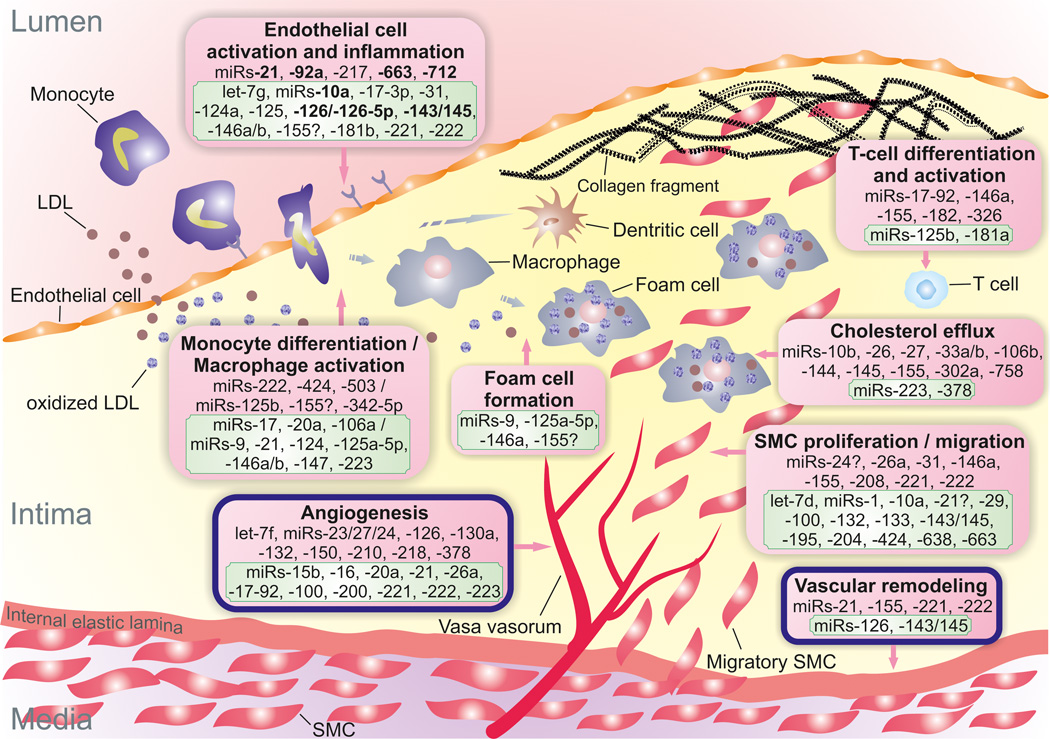
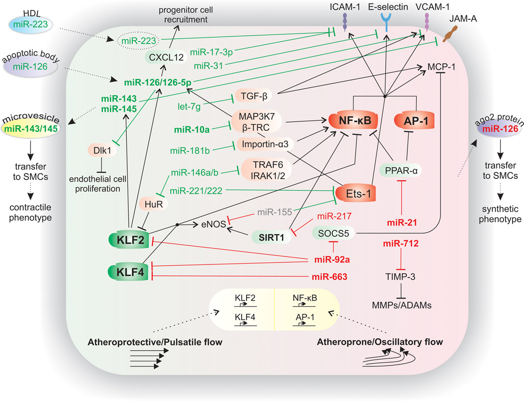
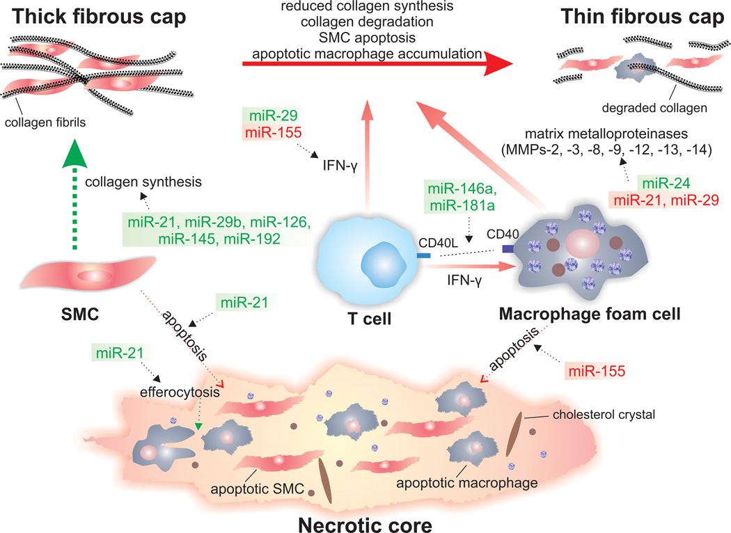
Atherosclerosis is a chronic immune-inflammatory disorder that integrates multiple cell types and a diverse set of inflammatory mediators. miRNAs are emerging as important post-transcriptional regulators of gene expression in most, if not all, vertebrate cells, and constitute central players in many physiological and pathological processes. Rapidly accumulating experimental studies reveal their key role in cellular and molecular processes related to the development of atherosclerosis. We review current evidence for the involvement of miRNAs in early atherosclerotic lesion formation and in plaque rupture and erosion. We conclude with a perspective on the clinical relevance, therapeutic opportunities, and future challenges of miRNA biology in understanding the pathogenesis of this complex disease.

Keywords: atherosclerosis; inflammation; miRNAs; shear stress; vulnerable plaque.


Figures

Figure 1
Figure 1. miRNAs implicated in atherosclerotic processes
Positive/atheroprotective (in green frame) or negative/atherogenic (in red frame) effects of miRNAs on atherosclerotic process are shown. Question marks next to miRNAs indicate controversial or contradictory evidence. miRNAs in bold are those reported to be regulated by blood flow/shear stress. LDL diffuses from the blood into the intima and undergoes oxidative modification. Oxidized LDL triggers the expression of leukocyte adhesion molecules by endothelial cells. The initial steps of atherosclerosis include adhesion of blood monocytes to the activated endothelium, their migration into the intima, their maturation into macrophages (or dendritic cells), and their uptake of lipid yielding foam cells. Although fewer in number than macrophages, other leukocyte subsets, such as T cells, also enter the intima and regulate cellular and humoral immune responses. Lesion progression involves the proliferation and migration of SMCs into the intima, as well as increased extracellular matrix protein synthesis, including collagen. Advanced lesions also exhibit intraplaque neovascularization and outward remodeling. Abbreviations: LDL, low-density lipoprotein; SMC, smooth muscle cell.
Figure 2
Figure 2. Endothelial miRNAs implicated in atherosclerosis
Established miRNAs that regulate, either in an atheroprotective (shown in green) or an atherogenic (shown in red) manner, inflammatory pathways in the endothelium and the impact of shear stress on these pathways are depicted. Arrows between miRNAs and mechanistic processes indicate stimulation of the processes by the corresponding miRNA. Blunted arrows indicate inhibition. miRNAs (and other factors) in bold are those shown to be regulated by blood flow/shear stress. Transcription factors KLF2 and KLF4 mediate the anti-inflammatory effects of high shear stress (atheroprotective/pulsatile flow) by inducing eNOS. In contrast, low shear stress (atheroprone/oscillatory flow) induces the activation of proinflammatory NF-κB and AP-1, thus promoting endothelial cell dysfunction and activation. The increased expression of the downstream target genes of these transcription factors, such as VCAM-1, ICAM-1, E-selectin, and MCP-1, promotes the attachment and infiltration of lipid and inflammatory cells into the subendothelial space. Abbreviations: ADAMs, a disintegrin and metalloproteinases; AP-1, activator protein-1; β-TRC, β-transducin repeat-containing gene; Dlk1, delta-like 1 homolog; eNOS, endothelial nitric oxide synthase; ICAM-1, intercellular adhesion molecule-1; IRAK1/2, interleukin-1receptor-associated kinase 1/2; KLF, Krüppel-like factor 2; JAM-A, junctional adhesion molecule-A; MAP3K7, mitogen-activated protein kinase kinase kinase 7; MCP-1, monocyte chemotactic protein-1; MMPs, matrix metalloproteinases; NF-κB, nuclear factor-κB; PPAR-α, peroxisome proliferator-activated receptor-α; SMC, smooth muscle cell; SIRT1, silent information regulator 1; SOCS5, suppressor of cytokine signaling 5; TIMP-3, tissue inhibitor of metalloproteinase-3; TGF-β, transforming growth factor-β; and TRAF6, tumor necrosis factor receptor-associated factor 6; VCAM-1, vascular cell adhesion molecule-1.
Figure 3
Figure 3. miRNAs implicated in atherosclerotic plaque destabilization
The potential involvements of miRNAs in molecular and cellular mechanisms associated with atherosclerotic plaque instability are illustrated. Green miRNAs indicate those with stabilizing properties, while red miRNAs indicates those with destabilizing properties. The advanced atherosclerotic plaque consists of a fibrous cap rich in SMCs and collagen. The gradual loss of SMCs by apoptosis and the increased activity of matrix-degrading enzymes result in a fragile and rupture-prone fibrous cap. T cells produce proinflammatory mediators, such as IFN-γ and CD40L, which contribute to the amplification of local inflammation and plaque instability. Moreover, IFN-γ inhibits the collagen synthesis by SMCs. CD40L interactions with CD40 lead further to the release and activation of MMPs from activated macrophages. Apoptotic macrophages and SMCs, as well as extracellular lipid derived from dead cells, can accumulate in advanced plaques leading to the formation of a soft, destabilizing necrotic core within the intima. Abbreviations: CD40L, CD40 ligand; IFN-γ, interferon-γ; MMPs, matrix metalloproteinases; SMC, smooth muscle cell.

References

    1. Bartel DP. MicroRNAs: target recognition and regulatory functions. Cell. 2009;136:215–233. - PMC - PubMed
    1. Suarez Y, et al. Cutting edge: TNF-induced microRNAs regulate TNF-induced expression of E-selectin and intercellular adhesion molecule-1 on human endothelial cells: feedback control of inflammation. J Immunol. 2010;184:21–25. - PMC - PubMed
    1. Zhu N, et al. Endothelial enriched microRNAs regulate angiotensin II-induced endothelial inflammation and migration. Atherosclerosis. 2011;215:286–293. - PubMed
    1. Sun HX, et al. Essential role of microRNA-155 in regulating endothelium-dependent vasorelaxation by targeting endothelial nitric oxide synthase. Hypertension. 2012;60:1407–1414. - PubMed
    1. Sun X, et al. MicroRNA-181b regulates NF-kappaB-mediated vascular inflammation. J Clin Invest. 2012;122:1973–1990. - PMC - PubMed

Publication types