Review
. 2015 Apr;21(4):233-44.
doi: 10.1016/j.molmed.2015.02.006. Epub 2015 Mar 11.

Autoimmune host-microbiota interactions at barrier sites and beyond

Affiliations
Review

Autoimmune host-microbiota interactions at barrier sites and beyond

William E Ruff et al. Trends Mol Med. 2015 Apr.

Abstract

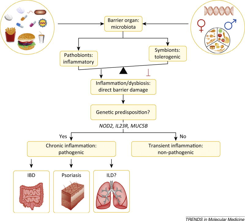
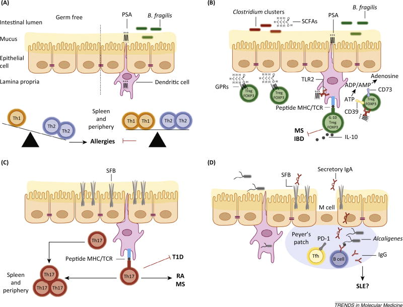
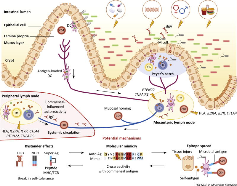
The microbiota is considered to be an important factor influencing the pathogenesis of autoimmunity at both barrier sites and internal organs. Impinging on innate and adaptive immunity, commensals exert protective or detrimental effects on various autoimmune animal models. Human microbiome studies of autoimmunity remain largely descriptive, but suggest a role for dysbiosis in autoimmune disease. Humanized gnotobiotic approaches have advanced our understanding of immune-commensal interactions, but little is known about the mechanisms in autoimmunity. We propose that, similarly to infectious agents, the microbiota mediates autoimmunity via bystander activation, epitope spread, and, particularly under homeostatic conditions, via crossreactivity. This review presents an overview of the current literature concluding with outstanding questions in this field.

Keywords: bystander activation; commensal; crossreactivity; epitope spread; microbiome; molecular mimicry.


Figures

Figure 1
Figure 1
Gut microbiota balance inflammation at barrier organs. Complex interactions between genetics, environment, and the microbiota shape the inflammatory status at barrier sites, and this impinges on autoinflammatory and autoimmune diseases. Diet, hygiene, antibiotics, pathogenic infections, and hormones shape the balance between pathobionts and symbionts at barrier organs. The barrier sites themselves, as well as the effects of each of these external factors on the barrier organs, are influenced by genetics. Increases in pathobionts or decreases in anti-inflammatory commensals favor aberrant immune interactions with these microbes, leading to dysbiosis and direct barrier damage. In genetically predisposed individuals, barrier damage is thought to instigate inflammatory bowel disease (IBD) in the gastrointestinal tract and psoriasis at cutaneous surfaces. The susceptible sites are determined by the colonization patterns of commensals. Other barrier sites might be equally affected by the microbiota, for example interstitial lung disease (ILD) in the lung. Individuals not genetically predisposed will have only transient or no inflammation with limited pathogenesis. Genes implicated in gut, skin, or lung barrier integrity are, for example, nucleotide-binding oligomerization domain-containing protein 2 (NOD2; polymorphic in IBD), interleukin-23 receptor (IL23R; polymorphic in psoriasis), and mucin 5B (MUC5B; polymorphic in ILD).
Figure 2
Figure 2
Microbiota influence CD4+ helper T (TH) cell subsets. (A) Germ-free animals have decreased numbers of CD4+ T cells in the periphery. The splenic CD4+ T cell population of germ-free animals is skewed towards TH2 cells over TH1 cells, favoring allergic diseases. This phenotype is reversed by monocolonization with the zwitterionic polysaccharide-A (PSA)-producing Bacteroides fragilis (B. fragilis) or oral administration of PSA alone. This process is mediated through the recognition and presentation of PSA by dendritic cells (DCs) sampling the intestinal lumen. (B) In the gut, PSA signals through the innate toll-like receptor 2 (TLR2) mediating the expansion of CD39+ FOXP3+ interleukin-10+ (IL-10) Tregs. The ectonucleotidases CD39 in conjunction with CD73 promote degradation of ATP. PSA-induced Tregs have been shown to inhibit multiple sclerosis (MS) and colitis in animal models. Additional anti-inflammatory effects are elicited by bacterial production of short-chain fatty acids (SCFAs) that also dampen colitis. SCFAs promote the stabilization of Tregs by inhibiting histone deacetylases and triggering chemotactic G protein-coupled receptors (GPRs) on FOXP3+ Tregs. (C) Segmented filamentous bacteria (SFB) induce the differentiation of inflammatory CD4+ TH17 cells through direct interactions with intestinal epithelial cells and MHC class II-dependent antigen presentation of SFB antigens to CD4+ T cells by intestinal DCs. The induction of intestinal TH17 cells by SFB has been linked to inhibition of type 1 diabetes (T1D) and the induction of rheumatoid arthritis (RA) and MS in animal models. (D) SFB and Alcaligenes species promote the antigen-specific and antigen-independent production of immunoglobulin A (IgA) in the intestinal lumen and in the periphery. The gut microbiota influences the production of T cell dependent immunoglobulin (Ig) production likely through PD-1+ follicular TH cells (TFH) that are known to instruct B cells to produce antigen-specific Ig. This process could influence systemic lupus erythematosus (SLE) or other autoantibody-mediated autoimmune diseases.
Figure 3
Figure 3
The gut microbiota impinges on non-gut autoimmunity. The gut microbiota balances the development of organ-specific and systemic autoimmunity in animal models. Dietary, antibiotic, genetic, hormonal, and hygienic factors affect the composition of the intestinal microbiota. Altered microbial community composition or function (dysbiosis) influences immune homeostasis locally and systemically. Genetic susceptibility (particularly via HLA haplotypes and genes regulating inflammation), combined with barrier disruption and dysbiosis, will increase the propensity of antigen-presenting cells [e.g., dendritic cells (DCs) or B cells] to take up antigen at different sites (lamina propria and Peyer’s patches), become activated, and present antigen to cognate T cells in secondary lymphoid organs. Follicular TH cells (TFH; shown as Tfh) cells help B cells in the germinal centers of lymph nodes or Peyer’s patches to produce different classes of antibodies, such as secretory IgA (sIgA) in the gut and IgG and IgA in the periphery. Activated immune cells traffic from the intestine to the mesenteric lymph nodes where they become imprinted with intestinal-homing markers. Some cells will also circulate systemically entering peripheral lymph nodes and target tissues. The development of autoreactive lymphocytes is proposed to occur due to three mechanisms: bystander effects, molecular mimicry, and epitope spreading. These mechanisms are not mutually exclusive and can also affect barrier organ autoimmunity. Each of these three mechanisms applied to commensals could be implicated in the development of non-barrier organ autoimmune diseases, for example type 1 diabetes (T1D), rheumatoid arthritis (RA), multiple sclerosis (MS), or systemic lupus erythematosus (SLE), similarly to transient autoimmune syndromes induced by pathogenic infections affecting for instance the joints (rheumatic fever) or nervous system (Guillan–Barré syndrome). Without genetic susceptibility, the proposed anti-commensal responses would be limited and lead to no overt autoimmunity. In a genetically predisposed host, several scenarios can occur depending on the genetics and pathobiont/ symbiont balance. Dysbiosis could be sustained by a genetic predisposition that leads to innate or B cell hyper-responsiveness, for example via polymorphisms in protein tyrosine phosphatase 22 (PTPN22) or tumor necrosis factor α-induced protein 3 (TNFAIP3). Without overt dysbiosis, antigen-specific recognition of commensals could lead to autoimmunity via crossreactivity if HLA polymorphisms or genetically encoded defects in Tregs or T cell homeostasis are present, for example in interleukin-2 receptor a (IL2RA), interleukin-7 receptor (IL7R), or cytotoxic T lymphocyte-associated protein 4 (CTLA4).

References

    1. Belkaid Y, Naik S. Compartmentalized and systemic control of tissue immunity by commensals. Nat. Immunol. 2013;14:646–653. - PMC - PubMed
    1. Brestoff JR, Artis D. Commensal bacteria at the interface of host metabolism and the immune system. Nat. Immunol. 2013;14:676–684. - PMC - PubMed
    1. Sommer F, Backhed F. The gut microbiota - masters of host development and physiology. Nat. Rev. Microbiol. 2013;11:227–238. - PubMed
    1. Maynard CL, et al. Reciprocal interactions of the intestinal microbiota and immune system. Nature. 2012;489:231–241. - PMC - PubMed
    1. Belkaid Y, Hand TW. Role of the microbiota in immunity and inflammation. Cell. 2014;157:121–141. - PMC - PubMed

Publication types