. 2015 Mar 23;32(6):719-30.
doi: 10.1016/j.devcel.2015.02.014. Epub 2015 Mar 12.

Notum is required for neural and head induction via Wnt deacylation, oxidation, and inactivation

Affiliations

Notum is required for neural and head induction via Wnt deacylation, oxidation, and inactivation

Xinjun Zhang et al. Dev Cell. .

Abstract

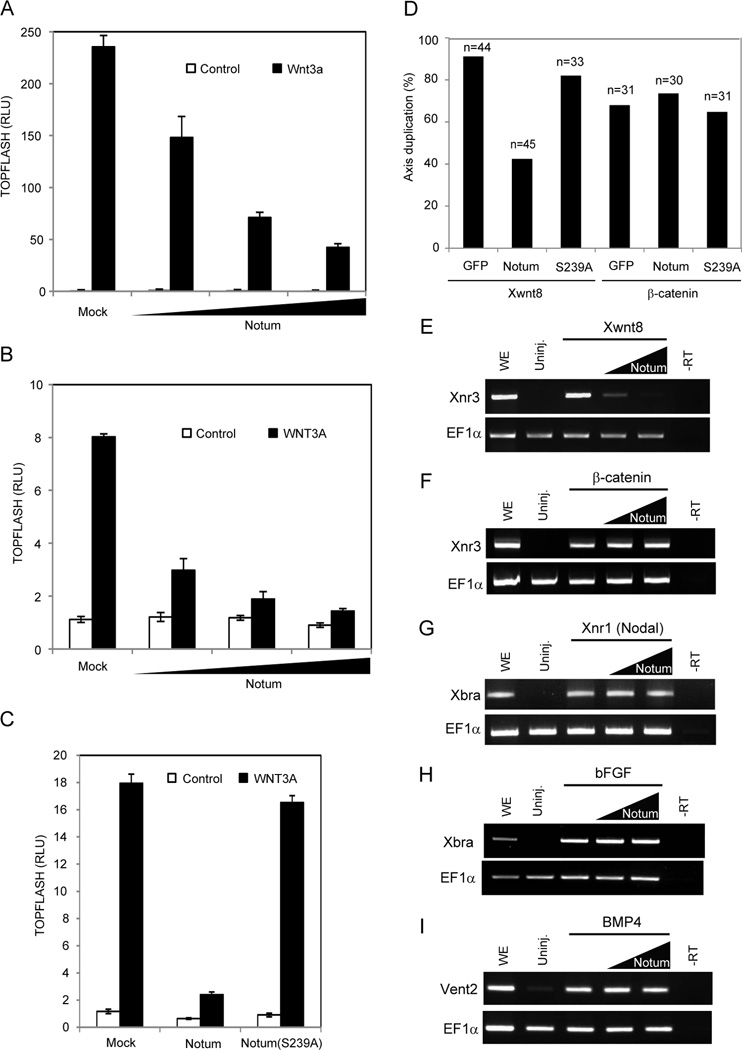
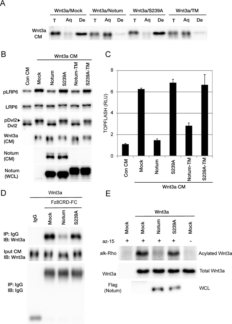
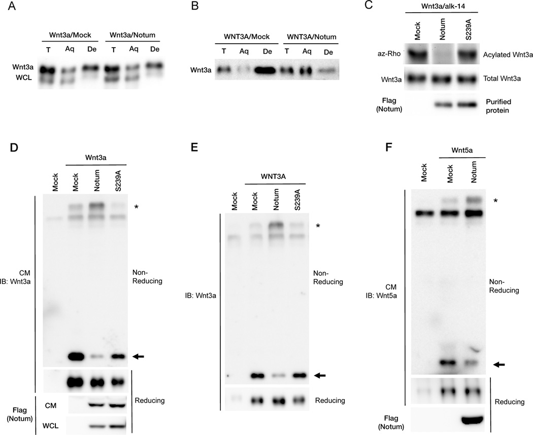
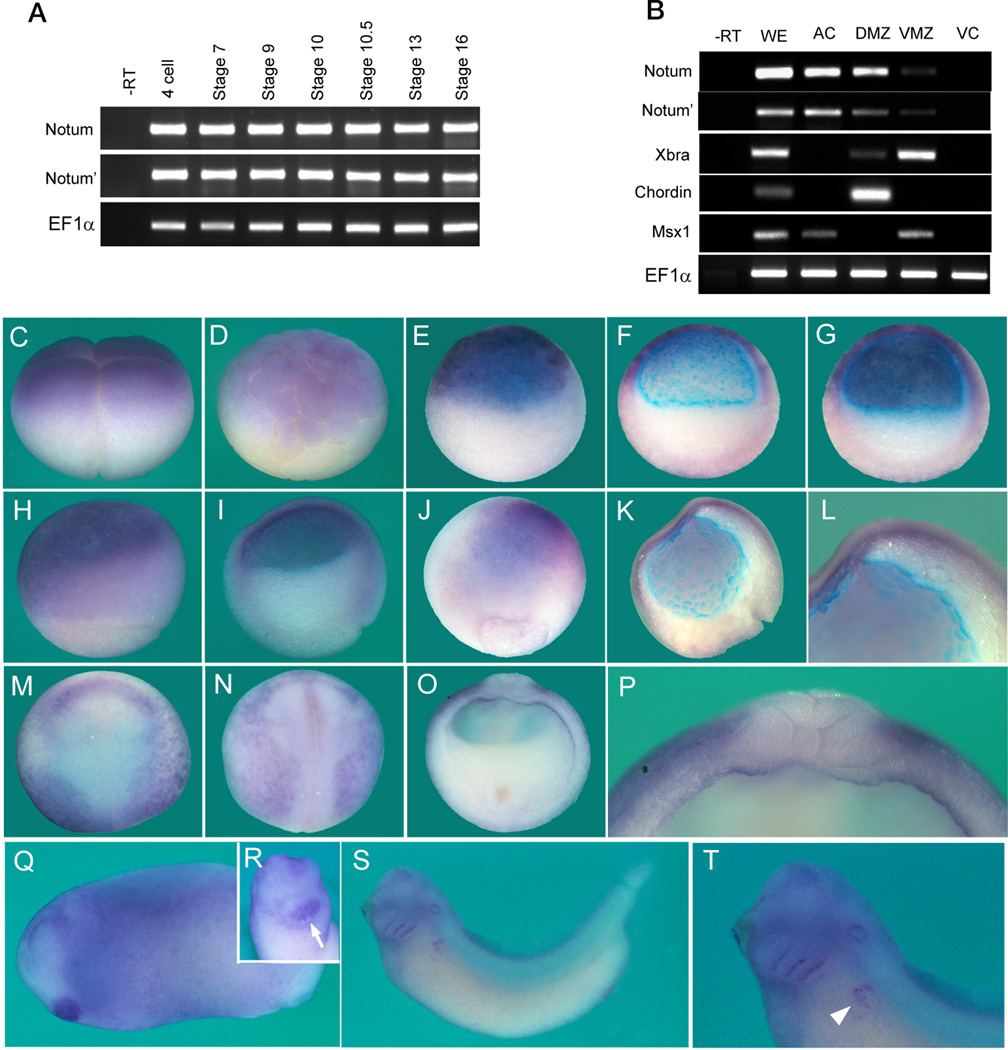
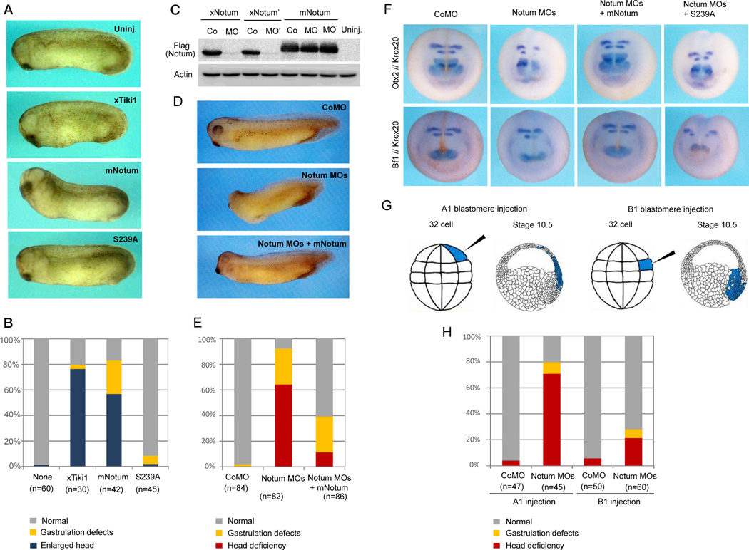
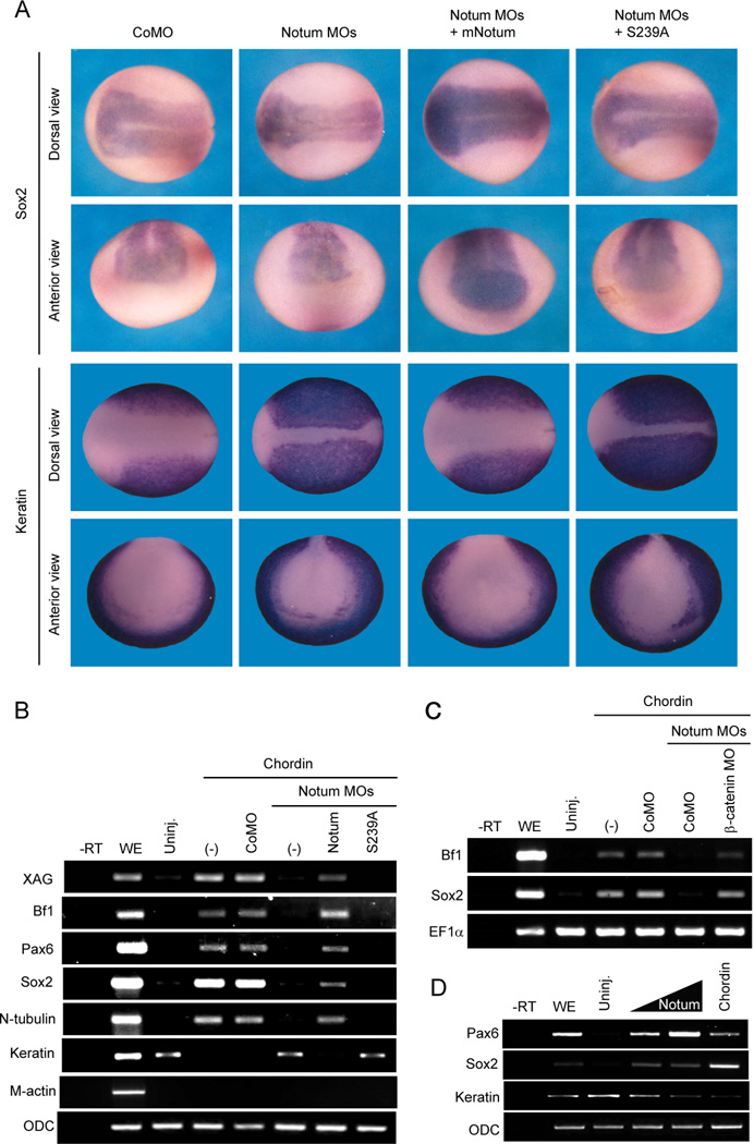
Secreted Wnt morphogens are essential for embryogenesis and homeostasis and require a lipid/palmitoleoylate modification for receptor binding and activity. Notum is a secreted Wnt antagonist that belongs to the α/β hydrolase superfamily, but its mechanism of action and roles in vertebrate embryogenesis are not fully understood. Here, we report that Notum hydrolyzes the Wnt palmitoleoylate adduct extracellularly, resulting in inactivated Wnt proteins that form oxidized oligomers incapable of receptor binding. Thus, Notum is a Wnt deacylase, and palmitoleoylation is obligatory for the Wnt structure that maintains its active monomeric conformation. Notum is expressed in naive ectoderm and neural plate in Xenopus and is required for neural and head induction. These findings suggest that Notum is a prerequisite for the "default" neural fate and that distinct mechanisms of Wnt inactivation by the Tiki protease in the Organizer and the Notum deacylase in presumptive neuroectoderm orchestrate vertebrate brain development.


Figures

Figure 1
Figure 1. Notum antagonizes Wnt signaling in mammalian cells and Xenopus embryos
(A) Notum inhibited TOPFLASH induced by Wnt3a in HEK293T cells. Increasing doses of a Notum expression vector were co-transfected with an expression vector for Wnt3a. (B) Notum inhibited TOPFLASH induced by the recombinant WNT3A protein. HEK293T cells transfected with increasing doses of the Notum expression vector were incubated with the WNT3A protein. (C) Notum(S239A) lacked the ability to inhibit TOPFLASH induced by the WNT3A protein. (D) Notum inhibited axis duplication induced by Xenopus Wnt8 but not β-catenin, while Notum(S239A) did not inhibit axis duplication induced by either Wnt8 or β-catenin. n: embryos examined. (E to I) Notum in animal pole explants inhibited expression of Xnr3 induced by Wnt8 (E), but not by β-catenin (F), nor did it inhibit Xbra expression induced by Nodal/Xnr1 (G) or bFGF (H), or Vent2 expression induced by BMP4 (I). EF1α, a loading control; uninj., uninjected embryos; WE, whole embryos; -RT, whole embryos without the reverse transcriptase. Two doses of Notum mRNAs were injected. Error bars (A to C) represent SD of triplicated experiments. See also Figure S1.
Figure 2
Figure 2. Notum causes Wnt deacylation and inactivation
(A) The Wnt3a protein modified by Notum became hydrophilic in the Triton X-114 detergent-aqueous phase separation assay. Wnt3a from mock or Notum(S239A)-expressing cells was hydrophobic and partitioned in the detergent (De) phase, but Wnt3a from Notum- or Notum-TM-expressing cells partitioned in the aqueous (Aq) phase. T, total input. (B) Wnt3a CM from Notum- or Notum-TM-expressing cells was inactive and induced minimal phosphorylation of LRP6 and Dvl2 in mouse embryonic fibroblast cells, whereas Wnt3a CM from mock, Notum(S239A)- or Notum(S239A)-TM-expressing cells induced phosphorylation of LRP6 and Dvl2. Note that Wnt3a from Notum- or Notum-TM-expressing cells exhibited slightly faster migration. WCL: whole cell lysates. (C) Wnt3a CM from Notum or Notum-TM-expressing cells induced minimal TOPFLASH in HEK293T cells, whereas Wnt3a CM from mock, Notum(S239A)- or Notum(S239A)-TM-expressing cells induced TOPFLASH. Error bars represent SD of triplicated experiments. (D) Wnt3a secreted from Notum-expressing cells exhibited minimal binding to mFz8CRD-IgG, whereas Wnt3a secreted from mock or Notum(S239A)-expressing cells exhibited binding. (E) Notum but not Notum(S239A) reduced Wnt3a acylation when they were coexpressed in HEK293T cells. See also Figure S2.
Figure 3
Figure 3. Notum is likely a Wnt deacylase acting extracellularly and causes Wnt3a and Wnt5a to form oxidized oligomers
(A) Notum had minimal effects on hydrophobicity of Wnt3a from WCL in the detergent-aqueous phase separation assay. (B) Recombinant WNT3A proteins lost hydrophobicity after incubation with Notum-expressing cells. (C) Notum reduced Wnt3a acylation in vitro. Purified metabolically labeled Wnt3a proteins were incubated with mock or purified Notum or Notum(S239A) proteins in vitro. (D) Wnt3a secreted from Notum- but not Notum(S239A)-expressing cells formed oxidized oligomer. Wnt3a CM from mock, Notum- or Notum(S239A)-expressing cells was analyzed by non-reducing or reducing SDS-PAGE. Wnt3a monomers and oxidized oligomers were labeled by an arrow and asterisk, respectively. (E) Recombinant WNT3A proteins formed oxidized oligomers upon incubation with Notum-, but not mock or Notum(S239A)-expressing cells. (F) Wnt5a proteins secreted from Notum-expressing cells formed oxidized oligomers. See also Figure S2.
Figure 4
Figure 4. Notum expression patterns during Xenopus embryogenesis
(A and B) RT-PCR revealed that Notum is maternally and zygotically expressed throughout Xenopus embryogenesis (A), and is enriched animally and dorsally at the stage 10.5. AC: animal caps; DMZ: dorsal marginal zone; VMZ: ventral marginal zone; VC, vegetal caps; Xbra: a pan-mesodermal marker; Chordin: a dorsal marker; Msx1: an animal and ventral marker. (C to T) Whole mount in situ hybridization for Notum/Notum’ expression, showing lateral view at 4-cell and stage 6.5 (C and D); lateral and bisected view at stage 8.5 (E and F); bisected view at stage 9.5 (G); lateral and bisected view at stage 10.5, with dorsal on right (H and I); dorsal and bisected view at the stage 11 (J and K) with an enlarged anterior view showing stronger expression anteriorly (L); dorso-anterior (M), dorsal (N, anterior on top), and cross-section (O, dorsal on top) views at stage 15 with an enlarged dorsal view (P); lateral view at the tailbud stage (Q), with an enlarged anterior view indicating expression in the cement gland (R, arrow); lateral view at the early tadpole stage (S), with an enlarged view indicating expression in the pronephros region (T, arrowhead). The blue color at the blastocoel surface in bisections was non-specific. See also Figures S3.
Figure 5
Figure 5. Notum is required for anterior development in Xenopus embryos
(A and B) Dorsal injection of Notum mRNA, but not Notum(S239A), induced an enlarged head similar to that induced by Tiki1 mRNA, and resulting phenotypes were tabulated. (C) The Notum MO and the Notum’ MO inhibited protein synthesis from Xenopus Notum (xNotum) or Notum’ (xNotum’) but not mouse Notum (mNotum) mRNAs. Co, MO, and MO’ indicate control MO and MOs against xNotum and xNotum’, respectively. (D and E) Dorso-animal injection of the two Notum MOs together caused anterior defects that were rescued by mNotum mRNA, and resulting phenotypes were tabulated. (F) The Notum MOs suppressed expression of forebrain markers Otx2 and Bf1 and a hindbrain marker Krox20 at stage 17. See also Table S1. (G) Illustration of the MO-injected A1 and B1 blastomeres at 32-cell stage and their descendent tissues at stage 10.5. (H) The Notum MOs injected into the A1 blastomeres caused anterior defects in more embryos (69%) than those injected into the B1 blastomeres (22%). See also Figure S4, S5, Table S2 and S3.
Figure 6
Figure 6. Notum is required for neural induction in embryos and animal explants
(A) The Notum MOs reduced the expression domain of Sox2, a pan-neural marker, and expanded the expression domain of cytokeratin, an epidermal marker, and the reciprocal changes were rescued by mNotum, but not mNutum(S239A), mRNA. See also Table S4. (B) The Notum MOs suppressed expression of neural markers induced by injection of the Chordin mRNA, and restored cytokeratin expression that was inhibited by Chordin. The effect of Notum MOs was rescued by mNotum, but not mNotum(S239A), mRNA. XAG, a cement gland marker; Bf1 and Pax6, anterior neural markers; Sox2, a pan-neural marker; N-tubulin, a neuronal marker; Keratin, an epidermal marker; M-Actin, a mesodermal marker; ODC, a loading control. (C) Chordin induced Bf1 and Sox2 expression when the Notum MOs and a β-catenin MO were co-injected. (D) Injection of Notum mRNA, like that of Chordin mRNA, induced expression of neural markers Sox2 and Pax6 and suppressed that of an epidermal marker, cytokeratin, in animal cap explants. See also Figure S6.

References

    1. Alexandre C, Baena-Lopez A, Vincent JP. Patterning and growth control by membrane-tethered Wingless. Nature. 2014;505:180–185. - PMC - PubMed
    1. Barrott JJ, Cash GM, Smith AP, Barrow JR, Murtaugh LC. Deletion of mouse Porcn blocks Wnt ligand secretion and reveals an ectodermal etiology of human focal dermal hypoplasia/Goltz syndrome. Proceedings of the National Academy of Sciences of the United States of America. 2011;108:12752–12757. - PMC - PubMed
    1. Beckett K, Franch-Marro X, Vincent JP. Glypican-mediated endocytosis of Hedgehog has opposite effects in flies and mice. Trends in cell biology. 2008;18:360–363. - PubMed
    1. Biechele S, Cox BJ, Rossant J. Porcupine homolog is required for canonical Wnt signaling and gastrulation in mouse embryos. Developmental biology. 2011;355:275–285. - PubMed
    1. Cantu JA, Flowers GP, Topczewski J. Notum homolog plays a novel role in primary motor innervation. The Journal of neuroscience. 2013;33:2177–2187. - PMC - PubMed

Publication types

MeSH terms

Associated data