. 2015 Apr 2;16(4):367-72.
doi: 10.1016/j.stem.2015.02.005. Epub 2015 Mar 12.

Lymphoid regeneration from gene-corrected SCID-X1 subject-derived iPSCs

Affiliations

Lymphoid regeneration from gene-corrected SCID-X1 subject-derived iPSCs

Tushar Menon et al. Cell Stem Cell. .

Abstract

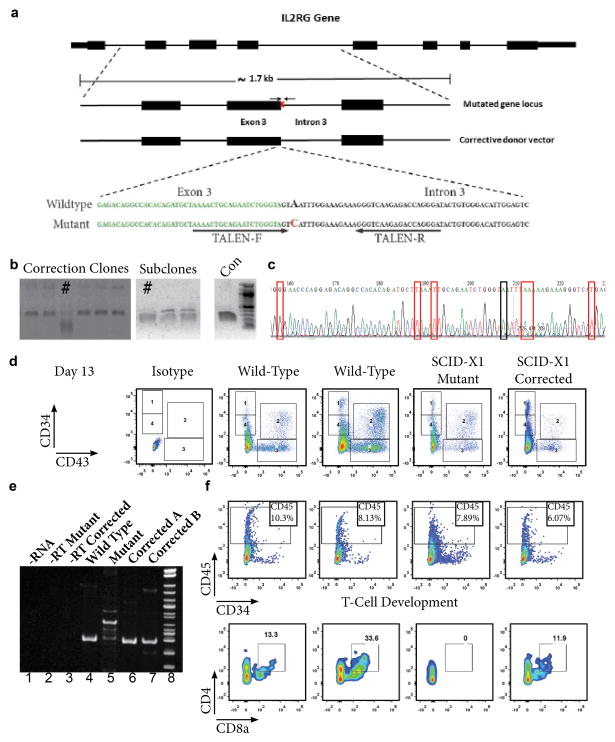
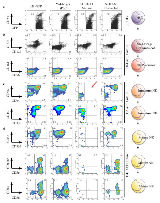
X-linked Severe Combined Immunodeficiency (SCID-X1) is a genetic disease that leaves newborns at high risk of serious infection and a predicted life span of less than 1 year in the absence of a matched bone marrow donor. The disease pathogenesis is due to mutations in the gene encoding the Interleukin-2 receptor gamma chain (IL-2Rγ), leading to a lack of functional lymphocytes. With the leukemogenic concerns of viral gene therapy there is a need to explore alternative therapeutic options. We have utilized induced pluripotent stem cell (iPSC) technology and genome editing mediated by TALENs to generate isogenic subject-specific mutant and gene-corrected iPSC lines. While the subject-derived mutant iPSCs have the capacity to generate hematopoietic precursors and myeloid cells, only wild-type and gene-corrected iPSCs can additionally generate mature NK cells and T cell precursors expressing the correctly spliced IL-2Rγ. This study highlights the potential for the development of autologous cell therapy for SCID-X1 subjects.


Figures

Figure 1
Figure 1. TALEN Mediated Gene Correction and Lymphoid Differentiation of SCID-X1 iPSC
a) Schematic representation of the IL2Rγ gene, the endogenous target locus of our patient-specific mutation and the corrective donor vector used. The sequence of the patient-specific IL2Rγ splice-site target mutation and the corrective donor vector sequence are also shown below. The point mutation causing the alteration of this exon/intron consensus splice site is indicated in red. Exon 3 sequence immediately preceding the splice site is denoted by green, bold bases. TALEN binding sites are indicated by black arrows. b) Identification and isolation of corrected iPSCs through single-cell clonal amplification and screening of the PCR products with an XmaI restriction digest that is specific to the correction event. Corrected clones and subclones are identified by #. c) Chromatogram of the corrected iPSC clone indicated in Fig. 1b, as verified by sequencing. The red boxes indicate each of the silent mutations that were introduced to abolish TALEN activity on the corrective or corrected DNA sequences. Black box indicates the corrected disease-causing base. d) Comparative analysis of FACS data from wild type, SCID-X1 mutant and SCID-X1 corrected iPSC. Data shows CD34 and CD43 expression at Day 13 of differentiation. Isotype controls are included in the left panel. e) RT-PCR analysis of RNA extracted from T-cell precursors in the floating fraction generated from wild type, patient-derived SCID-X1 mutant iPSC or SCID-X1 corrected iPSC clones. f) FACS analysis of the floating fraction of cells co-cultured on OP9-DL feeders from two independent wild type, the SCID-X1 mutant and SCID-X1 gene corrected iPSC line. CD45+ and CD45+/CD4+/CD8a+ positive populations are indicated.
Figure 2
Figure 2. Generation of Mature NK Cells
FACS analysis of dissociated cells from H1-Embryonic Stem Cells (ESC), Wild type iPSC, SCID-X1 Mutant iPSC and SCID-X1 Gene-corrected iPSC for the antigens indicated on the left at the following time points; a) Embryoid Bodies (EB) at Day 15; b) NK Lineage committed cells/NK precursors at Day 14 of AFT culture; c) Immature NK cells at Day 33 of AFT culture. The red arrow highlights the lack of CD56/CD94+ cells in the mutant iPSC and d) mature NK cells at Day 41 of AFT culture. Cell percentages are indicated at the corner of each gate.

Comment in

References

    1. Aiuti A, Biasco L, Scaramuzza S, Ferrua F, Cicalese MP, Baricordi C, Dionisio F, Calabria A, Giannelli S, Castiello MC, et al. Lentiviral hematopoietic stem cell gene therapy in patients with Wiskott-Aldrich syndrome. Science. 2013;341:1233151. - PMC - PubMed
    1. Bedell VM, Wang Y, Campbell JM, Poshusta TL, Starker CG, Krug RG, Tan WF, Penheiter SG, Ma AC, Leung AYH, et al. In vivo genome editing using a high-efficiency TALEN system. Nature. 2012;491:114–U133. - PMC - PubMed
    1. Biffi A, Montini E, Lorioli L, Cesani M, Fumagalli F, Plati T, Baldoli C, Martino S, Calabria A, Canale S, et al. Lentiviral hematopoietic stem cell gene therapy benefits metachromatic leukodystrophy. Science. 2013;341:1233158. - PubMed
    1. Biron CA, Nguyen KB, Pien GC, Cousens LP, Salazar-Mather TP. Natural killer cells in antiviral defense: function and regulation by innate cytokines. Annual review of immunology. 1999;17:189–220. - PubMed
    1. Boch J, Scholze H, Schornack S, Landgraf A, Hahn S, Kay S, Lahaye T, Nickstadt A, Bonas U. Breaking the code of DNA binding specificity of TAL-type III effectors. Science. 2009;326:1509–1512. - PubMed

Publication types

MeSH terms