. 2015 Aug;35(6):785-95.
doi: 10.1007/s10571-015-0172-z. Epub 2015 Mar 14.

The Internalization of Neurotensin by the Low-Affinity Neurotensin Receptors (NTSR2 and vNTSR2) Activates ERK 1/2 in Glioma Cells and Allows Neurotensin-Polyplex Transfection of tGAS1

Affiliations

The Internalization of Neurotensin by the Low-Affinity Neurotensin Receptors (NTSR2 and vNTSR2) Activates ERK 1/2 in Glioma Cells and Allows Neurotensin-Polyplex Transfection of tGAS1

Alberto E Ayala-Sarmiento et al. Cell Mol Neurobiol. 2015 Aug.

Abstract

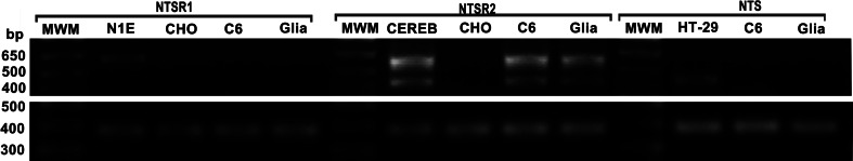
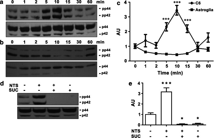
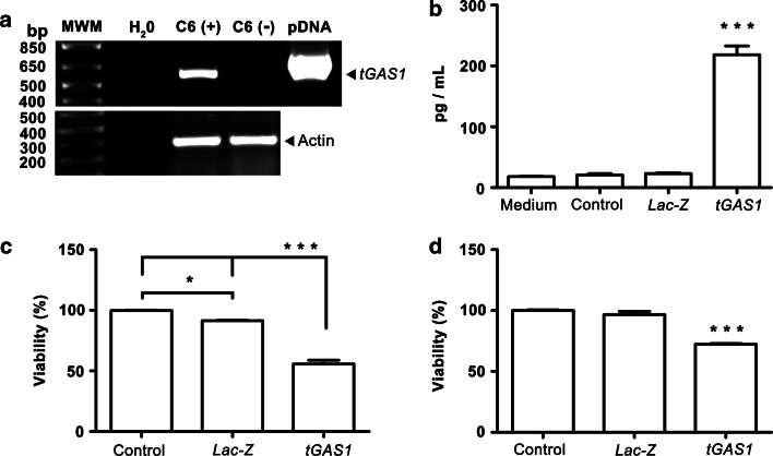
Glioblastoma is the most malignant primary brain tumor and is very resistant to treatment; hence, it has a poor prognosis. Neurotensin receptor type 1 (NTSR1) plays a key role in cancer malignancy and has potential therapeutic applications. However, the presence and function of neurotensin (NTS) receptors in glioblastoma is not clearly established. RT-PCR assays showed that healthy (non-tumor) astroglial cells and C6 glioma cells express NTSR2 and its isoform (vNTSR2) rather than NTSR1. In glioma cells, NTS promotes the phosphorylation of extracellular signal-regulated kinases 1/2 (ERK 1/2), an effect that was completely abolished by blocking the internalization of the NTS/NTSR complex. We demonstrated pharmacologically that the internalization is dependent on the activation of NTSR2 receptors and it was prevented by levocabastine, a NTSR2 receptor antagonist. The internalization of NTSR2 and vNTSR2 was further demonstrated by its ability to mediate gene transfer (transfection) via the NTS-polyplex system. Expression of reporter transgenes and of the pro-apoptotic soluble form of growth arrest specific 1 (tGAS1) was observed in glioma cells. A significant reduction on the viability of C6 cells was determined when tGAS1 was transfected into glioma cells. Conversely, astroglial cells could neither internalize NTS nor activate ERK 1/2 and could not be transfected by the NTS-polyplex. These results demonstrate that the internalization process of NTSR2 receptors is a key regulator necessary to trigger the activation of the ERK 1/2. Our data support a new internalization pathway in glioma C6 cells that involve NTSR2/vNTSR2, which can be used to selectively transfer therapeutic genes using the NTS-polyplex system.


Conflict of interest statement

The authors declare that they have no conflict of interest.

Figures

Fig. 1
Fig. 1
Expression of NTSR2 and vNTSR2 mRNAs in both murine enriched-astroglia cultures and murine glioma C6 cells. Representative gels showing the RT-PCR products for NTSR1 (620 bp), unspliced NTSR2 (600 bp), vNTSR2 (418 bp), or NTS (438 bp). Actin (377 bp) was detected in all cell extracts. N1E-115 cells, cerebellum, and HT29 cells were used as positive controls shown by the headings. CHO cells were used as negative control. MWM molecular weight markers
Fig. 2
Fig. 2
Internalization of the NTS-polyplex in glioma C6 cells. Representative micrographs of horizontal confocal sections of C6 cells incubated with the propidium iodide (PI)-labeled NTS-polyplex in the absence (ad) or in the presence of levocabastine (eh); enriched-astroglial primary cell cultures incubated with the propidium iodide (PI)-labeled NTS-polyplex in the absence (il) or in the presence of levocabastine (mp). Cytoskeleton, C6 cells were counterstained with phalloidin, whereas the astroglial cells were immunolabeled against GFAP (glial fibrillary acidic protein). Levo levocabastine. Nuclei were stained with Hoechst 33258. Arrowheads indicate PI fluorescent spots. Scale bar 25 µm
Fig. 3
Fig. 3
Role of NTSR2 and vNTSR2 receptors in the internalization-dependent activation of ERK 1/2 induced by NTS in glioma C6 cells. Representative western blot of glioma C6 cells (a) and astroglia (b) showing the phosphorylation levels of ERK 1/2 (upper panels) and total ERK 1/2 (lower panels) after stimulation with NTS (0.1 µM) from 1 to 60 min (headings). c Densitometric analysis of ERK 1/2 activation in glioma C6 (dots) and astroglial cells (squares). AU arbitrary units of the ratio between phosphorylated ERK 1/2 and total ERK 1/2. d Representative western blot of glioma C6 cells preincubated for 30 min without or with 0.45 M sucrose (suc) followed by a 10 min incubation with NTS (0.1 µM). Upper panel ERK 1/2 phosphorylation; lower panel total ERK 1/2. e Densitometric analysis of ERK 1/2 activation. Values represent the mean ± SEM of three independent experiments. * P < 0.05; ** P < 0.01; *** P < 0.001 compared with control, astroglial cells (c), or untreated cells (e). Densitometric analyses were analyzed by two-way repeated measures ANOVA (c) or One-way ANOVA (e) and then by post hoc Bonferroni test
Fig. 4
Fig. 4
Transfection of glioma C6 cells by the NTS-polyplex. Representative confocal micrographs of C6 cells (ac) and astroglial cells (df) 36 h after exposure to the NTS-polyplex carrying the pEGFP-N1 plasmid. Cytoskeleton C6 cells were counterstained with phalloidin, whereas the astroglial cells were immunolabeled against GFAP (glial fibrillary acidic protein). Scale bar 25 µm
Fig. 5
Fig. 5
Effect of tGAS1 expression on the viability of glioma C6 cells. a Representative gels showing the RT-PCR products for tGAS1 (604 bp) and actin (377 bp) 36 h after exposure to the NTS-polyplex carrying the pLenti6.3/TO/V5-tGAS1 plasmid. MWM molecular weight markers, C6 (+) transfected cells, C6 () untransfected cells, pDNA pLenti6.3/TO/V5-tGAS1 plasmid. b Protein levels of tGAS1 assessed by ELISA in conditioned medium. Medium DMEM F-12 K, control conditioned medium from untransfected cells, Lac-Z conditioned medium from Lac-Z transfected cells, tGAS1 conditioned medium from tGAS1 transfected cells. c Cell viability using the MTT colorimetric assay in C6 cells after transfection (36 h) with the plasmid pLenti6.3/TO/V5-tGAS1 or d exposure to conditioned media (36 h) from previously transfected C6 cells. Values represent the mean ± SEM of three independent experiments. * P < 0.05 and *** P < 0.001 compared with control, untreated cells. One-way ANOVA and then by post hoc Bonferroni test

References

    1. Ahn S, Shenoy SK, Wei H, Lefkowitz RJ (2004) Differential kinetic and spatial patterns of beta-arrestin and G protein-mediated ERK activation by the angiotensin II receptor. J Biol Chem 279:35518–35525 - PubMed
    1. Alifano M et al (2010) Neurotensin receptor 1 determines the outcome of non-small cell lung cancer. Clin Cancer Res 16:4401–4410 - PubMed
    1. Alvarez-Maya I, Navarro-Quiroga I, Meraz-Rios MA, Aceves J, Martinez-Fong D (2001) In vivo gene transfer to dopamine neurons of rat substantia nigra via the high-affinity neurotensin receptor. Mol Med 7:186–192 - PMC - PubMed
    1. Arango-Rodriguez ML et al (2006) Biophysical characteristics of neurotensin polyplex for in vitro and in vivo gene transfection. Biochim Biophys Acta 1760:1009–1020 - PubMed
    1. Benitez JA, Arregui L, Vergara P, Segovia J (2007) Targeted-simultaneous expression of Gas1 and p53 using a bicistronic adenoviral vector in gliomas. Cancer Gene Ther 14:836–846 - PubMed

Publication types

MeSH terms

LinkOut - more resources