. 2015 Aug;63(4):299-304.
doi: 10.1007/s00005-015-0335-0. Epub 2015 Mar 13.

Macrophage Phenotype in the Ocular Surface of Experimental Murine Dry Eye Disease

Affiliations

Macrophage Phenotype in the Ocular Surface of Experimental Murine Dry Eye Disease

In-Cheon You et al. Arch Immunol Ther Exp (Warsz). 2015 Aug.

Abstract

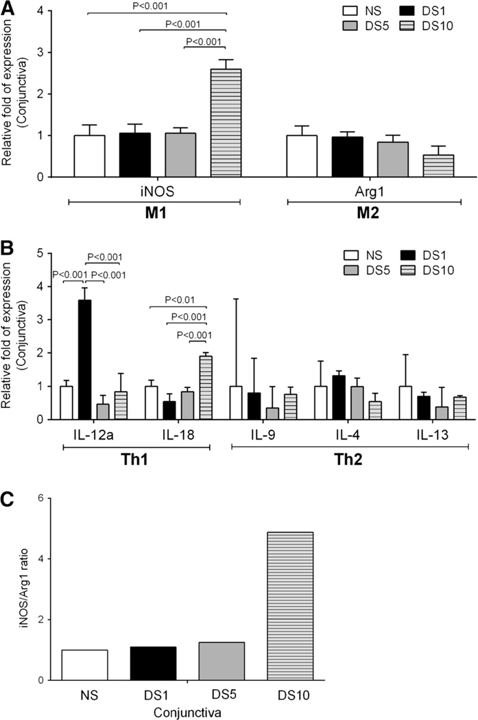
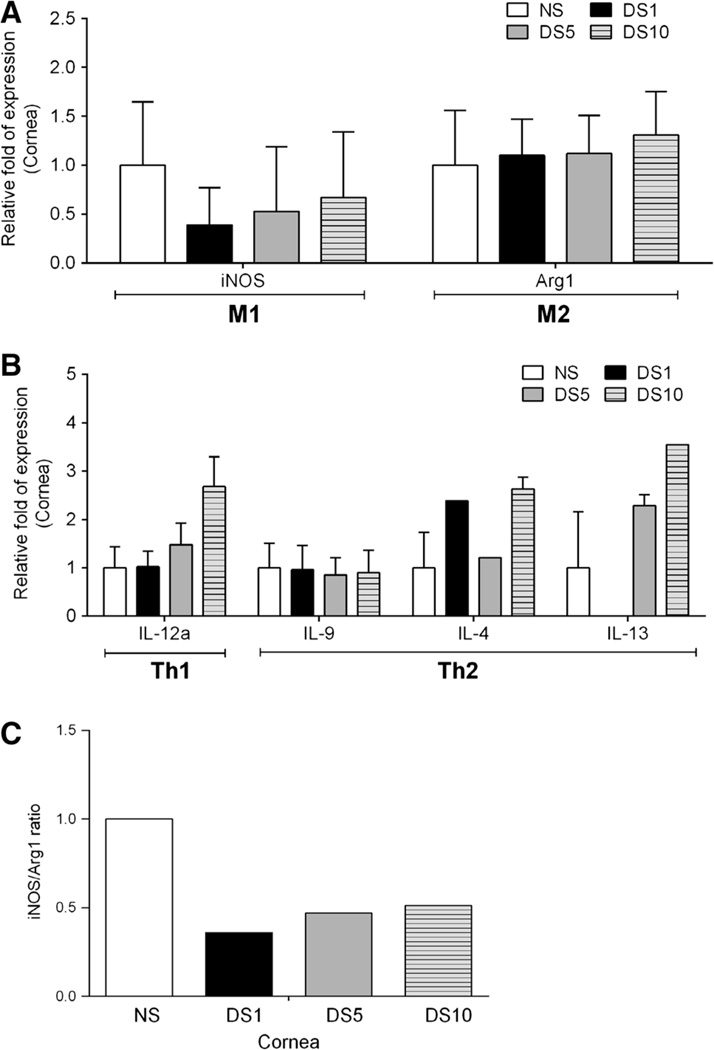
To evaluate the phenotype of macrophages in the cornea and conjunctiva of C57BL/6 mice with induced experimental dry eye. C57BL/6 mice exposed to desiccating stress (DS) were evaluated at 1, 5, and 10 days and C57BL/6 mice maintained in non-stressed environment were used as controls. Whole eyes and adnexa were excised for histology or used for gene expression analysis. Location and phenotype of macrophages infiltrating the cornea and conjunctiva was evaluated by immunofluorescence analysis. Quantitative polymerase chain reaction evaluated macrophage markers and T cell-related and inflammatory cytokine expression in cornea and conjunctiva. Immunofluorescence staining demonstrated that macrophages reside in the conjunctiva of control and dry eye mice and their number did not change with DS. Real-time RT-PCR demonstrated that the level of M1 macrophage marker, iNOS, increased prominently in the conjunctiva at DS 10 days. In contrast, there was a non-significant decrease of the M2 marker Arg1 with DS. The levels of inflammatory cytokine, IL-12a mRNA transcript in the conjunctiva increased significantly at DS1 and decreased at DS5, while levels of IL-18 were significantly increased at DS 10. Macrophages reside in the ocular surface tissues of C57BL/6 mice. Although the number of macrophages in the conjunctiva does not change, evidence of inflammatory M1 activation after desiccating stress was observed. Better understanding of phagocyte diversity and activation in dry eye disease provide a basis for the development of phagocyte-targeted therapeutic strategies.


Conflict of interest statement

Conflict of Interest All the authors declare that they have no conflict of interest.

Figures

Fig. 1
Fig. 1
Histologic analysis of macrophage infiltration in the ocular surface of C57BL/6 mice after desiccation stress. a Representative images showed macrophage localization [F4/80 (green)] in the cornea and conjunctiva by immunofluorescence staining with Hoechst (blue) as nuclear counterstaining. b Macrophage density in the conjunctiva. NS non-stressed, DS1 desiccating stress (DS) day 1, DS5 DS day 5, DS10 DS day 10
Fig. 2
Fig. 2
Quantitative gene analysis of mRNA transcripts in the conjunctiva of C57BL/6 mice after desiccation stress. a Relative fold of expression of M1 and M2 macrophage markers in conjunctiva of C57BL/6 mice at desiccating stress 1, 5, and 10 days. Bar graphs show mean ° SEM of one representative experiment with 7–10 samples/time point (experiment was repeated twice with similar results). One sample consisted of pooled right and left conjunctivas of the same animal. b Relative fold of expression of T cell-related cytokines and inflammatory cytokines. c iNOS to Arg1 ratio in the conjunctiva of C57BL/6 mice after desiccation stress. NS non-stressed, DS1 desiccating stress (DS) day 1, DS5 DS day 5, DS10 DS day 10, iNOS inducible nitric oxide synthase (NOS2), Arg1 arginase 1, IL-9 interleukin-9
Fig. 3
Fig. 3
Quantitative gene analysis of mRNA transcripts in the cornea of C57BL/6 mice after desiccation stress. a Relative fold of expression of M1 and M2 macrophage markers in cornea of C57BL/6 mice at desiccating stress 1, 5, and 10 days. Bar graphs show mean ± SEM of one representative experiment with 7–10 samples/time point (experiment was repeated twice with similar results). One sample consisted of pooled right and left cornea of the same animal. b Relative fold of expression of T cell-related cytokines and inflammatory cytokines. In results of IL-4 and IL-13, there was 1/5 less only sample to amplify at DS1, DS5, and DS10. c iNOS to Arg1 ratio in the cornea of C57BL/6 mice after desiccation stress. NS non-stressed, DS1 desiccating stress (DS) day 1, DS5 DS day 5, DS10 DS day 10, iNOS inducible nitric oxide synthase (NOS2), Arg1 arginase 1, IL-9 interleukin-9

References

    1. Biswas SK, Mantovani A. Macrophage plasticity and interaction with lymphocyte subsets: cancer as a paradigm. Nat Immunol. 2010;11:889–896. - PubMed
    1. Chiang CS, Chen FH, Hong JH, et al. Functional phenotype of macrophages depends on assay procedures. Int Immunol. 2008;20:215–222. - PubMed
    1. Cho DI, Kim MR, Jeong HY, et al. Mesenchymal stem cells reciprocally regulate the M1/M2 balance in mouse bone marrow-derived macrophages. Exp Mol Med. 2014;46:e70. - PMC - PubMed
    1. Coursey TG, Gandhi NB, Volpe EA, et al. Chemokine receptors CCR6 and CXCR3 are necessary for CD4(+) T cell mediated ocular surface disease in experimental dry eye disease. PLoS One. 2013;8:e78508. - PMC - PubMed
    1. De Paiva CS, Villarreal AL, Corrales RM, et al. Dry eye-induced conjunctival epithelial squamous metaplasia is modulated by interferon-gamma. Invest Ophthalmol Vis Sci. 2007;48:2553–2560. - PubMed

Publication types

LinkOut - more resources