Review
. 2015 Sep:81:134-43.
doi: 10.1016/j.nbd.2015.02.007. Epub 2015 Mar 12.

Neuroferritinopathy: From ferritin structure modification to pathogenetic mechanism

Affiliations
Review

Neuroferritinopathy: From ferritin structure modification to pathogenetic mechanism

Sonia Levi et al. Neurobiol Dis. 2015 Sep.

Abstract

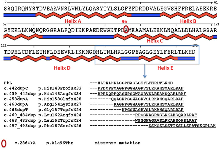
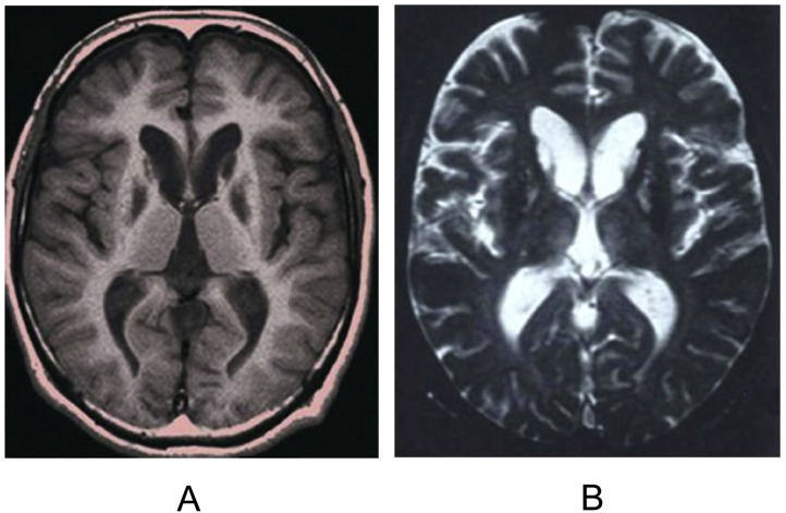
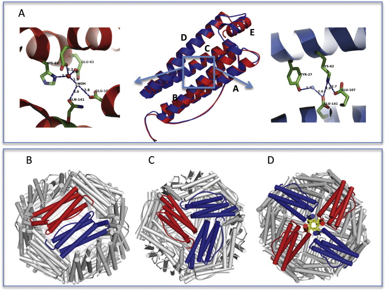
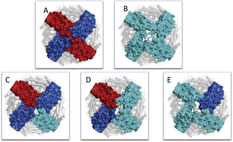
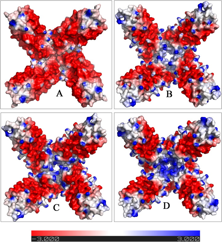
Neuroferritinopathy is a rare, late-onset, dominantly inherited movement disorder caused by mutations in L-ferritin gene. It is characterized by iron and ferritin aggregate accumulation in brain, normal or low serum ferritin levels and high variable clinical feature. To date, nine causative mutations have been identified and eight of them are frameshift mutations determined by nucleotide(s) insertion in the exon 4 of L-ferritin gene altering the structural conformation of the C-terminus of the L-ferritin subunit. Acting in a dominant negative manner, mutations are responsible for an impairment of the iron storage efficiency of ferritin molecule. Here, we review the main characteristics of neuroferritinopathy and present a computational analysis of some representative recently defined mutations with the purpose to gain new information about the pathogenetic mechanism of the disorder. This is particularly important as neuroferritinopathy can be considered an interesting model to study the relationship between iron, oxidative stress and neurodegeneration.

Keywords: Ferritin; Iron; Neurodegenerative disorder; Neuroferritinopathy; Oxidative damage.


Figures

Fig. 1
Fig. 1
Schematic representation of L ferritin secondary structure and sequence alignment of FtL and NF variants. Human L ferritin sequence is matched with a schematic diagram of protein secondary structure elements that include five α-helices segments (red in upper figure), conventionally named with letters from A to E, connected by four loop elements and of two additional loops at the N and C termini (shown in blue). The red circle marks the position of residue 96, mutated in the unique missense variant (p.Ala96Thr). The blue rectangle highlight the sequence interval, from residue 148 to 175, involved in the so far detected frameshift variants. Frameshift mutations observed in patients are responsible for aminoacidic changes at the C-terminus starting from different sequence position depending on the duplication starting point. In panel B the alignment of the frameshift altered regions compared with the wild type FtL sequence is shown. Dashes correspond to unchanged position while modified residues are underlined.
Fig. 2
Fig. 2
Brain MRI in Neuroferritinopathy. Brain MR images of a Japanese man carrying the FTL1 c469_484dup mutation. Axial section at the level of the basal ganglia in the patient at 35 years of age. A A T1-weighted image (TR 400 msec/ TE 14 msec) shows symmetrical hypointense signals in the head of the caudate nucleus and globus pallidus. B A T2-weighted image (TR 800 msec/TE 30 msec) shows hypointense changes in the lenticular nucleus. Hyperintense signals can be observed in the putamen and the head of the caudate nucleus. Reproduced from Ohta E, Takiyama Y. MRI findings in neuroferritinopathy. Neurol Res Int. 2012;2012:197438.
Fig. 3
Fig. 3
Three-dimensional structure of ferritin. Ferritin subunit is a four-helix bundle protein consisting of two couples of anti-parallel α-helices (A–B and C–D) connected by a long loop and a short C-terminal α-helix (E) important for 24-mer stabilization. Human L and H ferritin subunits share a 55% sequence identity and a remarkably similar 3D structure. A In central panel is shown a ribbon representation of superposed L (blue) and H (red) human ferritin subunits. Superposition of the two structures yielded a root mean square deviation (RMSD) of 0.5 Å, indicating an elevated level of structural similarity. The two subunits show a different residue arrangement in the center of four-helix bundle, corresponding to the blue box. Arrows departing from the box point to the expanded representation of the region in H-chain (panel left side) and L-chain (panel right side). In H chain, a ferroxidase center is formed by a set of iron coordinating residues, which are replaced by a salt bridge forming amino acids in the L chain. The functional protein is a 24-mer polymer made of variable proportion of H and L chains that assemble following a two-three- and four-fold symmetry. The assembled ferritin appears as a hollow spherical shell (panel B). Subunit interactions in the 24-mer generate 12 dimeric interfaces (a dimer formed by one H and one L chain is shown as an example in panel B), eight hydrophilic channels, each obtained from 3 subunits, at the three fold axes (panel C) and six hydrophobic channels, obtained from 4 subunits at the four-fold axes (panel D). Structures were obtained from Protein Data Bank (PDB code: 2FFX for L- chain, 1FHA for H-chain; Berman et al, 2000).
Fig. 4
Fig. 4
Molecular surface representation of ferritin subunits forming the 4-fold axis hydrophobic pore. A Representative wild type pore formed by two FtL (blue) and two FtH (red) chains. The wild type channel appears very narrow due to the presence of tightly packed hydrophobic residues. B Putative pore formed by 4 identical FtLPhe167SerfsX26 subunits (cyan). The opening of the pore is largely increased, altering the iron permeability of the ferritin shell. In patients carrying FtL mutation, two different L chain alleles (wild type and mutated), in addition to the wild type H chain, are expressed; therefore, mixed heteropolymers are likely to occur. Three putative combinations of FtL (blue), FtH (red) and FtLPhe167SerfsX26 (cyan) subunits, have been reconstructed in panels C, D, and E, increasing the proportion of mutated chain. The pore width increases proportionally to the number of variant chains participating in the tetramer assembly. (The structure of FtLPhe167SerfsX26 variant was obtained from Protein Data Bank. PDB code: 2KXU).
Fig. 5
Fig. 5
Electrostatic potential distribution of the inner surface of homopolymeric 4 fold axis tetramers. The tetrameric assembly of four identical subunits was reconstructed starting from the wild type FtL subunit (panel A) and the variants p.Arg154LysfsX27, His148ProfsX33 and His148ProfsX33 (panels B, C, D, respectively). In wild type FtL tetramer (panel A), a prevalence of negative charges (red surface) is noticeable and is required for the iron nucleation inside the cavity. All the analyzed variants show an increased distribution of neutral or positive charges (white to blue surface in panels B-C-D) around the four-fold channel, and this can be responsible for a repulsive effect on iron complex. In addition, the reconstruction of the four-fold tetramers showed, for all variants, a wider pore compared to wild type, likely to alter the ferritin shell permeability. The potential scale ranges from − 3kT/e to + 3kT/e from red to blue. The PyMol molecular visualization system (The PyMOL Molecular Graphics System, Schrödinger, LLC) was used for structure visualization, analysis, comparison and for preparation of images.

References

    1. Adzhubei I., Jordan D.M., Sunyaev S.R. Predicting functional effect of human missense mutations using PolyPhen-2. In: Haines Jonathan L., editor. Current Protocols in Human Genetics/Editorial Board. 2013. (Chapter 7, Unit7 20) - PMC - PubMed
    1. Arosio P., Levi S. Cytosolic and mitochondrial ferritins in the regulation of cellular iron homeostasis and oxidative damage. Biochim. Biophys. Acta. 2010;1800:783–792. - PubMed
    1. Baraibar M.A., Barbeito A.G., Muhoberac B.B., Vidal R. Iron-mediated aggregation and a localized structural change characterize ferritin from a mutant light chain polypeptide that causes neurodegeneration. J. Biol. Chem. 2008;283:31679–31689. - PMC - PubMed
    1. Baraibar M.A., Muhoberac B.B., Garringer H.J., Hurley T.D., Vidal R. Unraveling of the E-helices and disruption of 4-fold pores are associated with iron mishandling in a mutant ferritin causing neurodegeneration. J. Biol. Chem. 2010;285:1950–1956. - PMC - PubMed
    1. Baraibar M.A., Barbeito A.G., Muhoberac B.B., Vidal R. A mutant light-chain ferritin that causes neurodegeneration has enhanced propensity toward oxidative damage. Free Radic. Biol. Med. 2012;52:1692–1697. - PMC - PubMed

Publication types

Supplementary concepts

LinkOut - more resources