. 2015 Mar 30;25(7):868-78.
doi: 10.1016/j.cub.2015.01.061. Epub 2015 Mar 12.

A dynamin-actin interaction is required for vesicle scission during endocytosis in yeast

Affiliations

A dynamin-actin interaction is required for vesicle scission during endocytosis in yeast

Sarah E Palmer et al. Curr Biol. .

Abstract

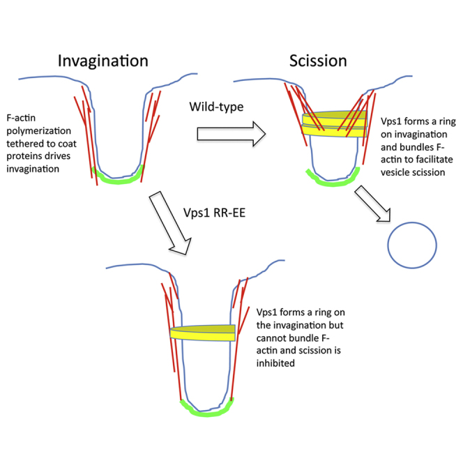
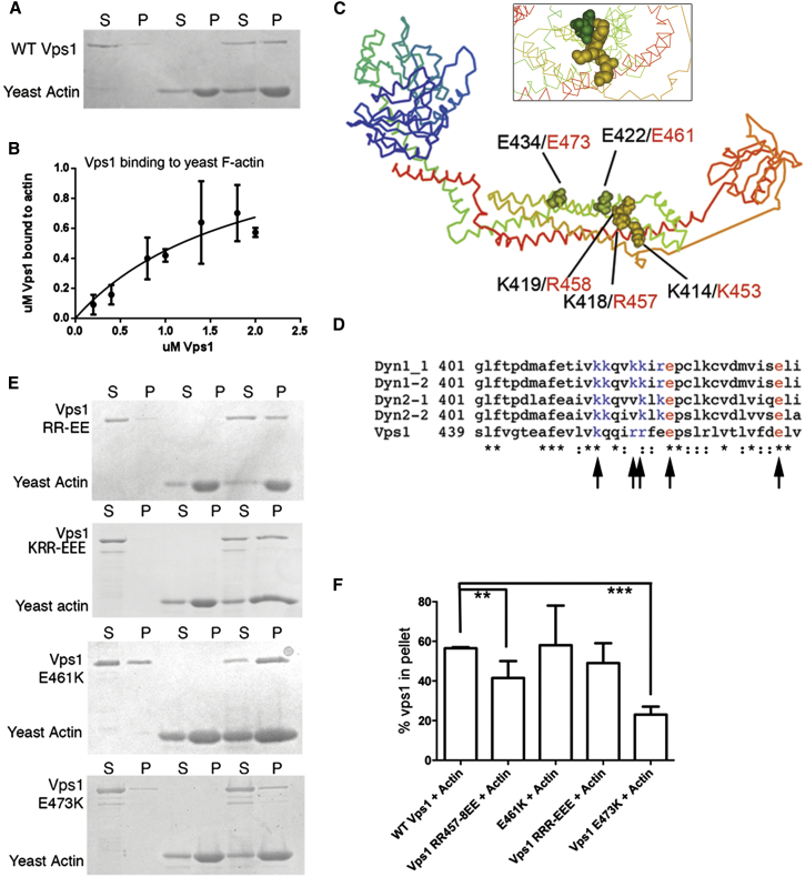
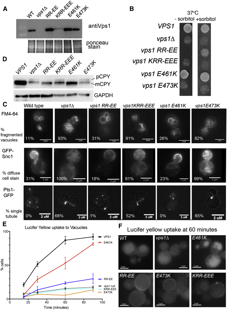
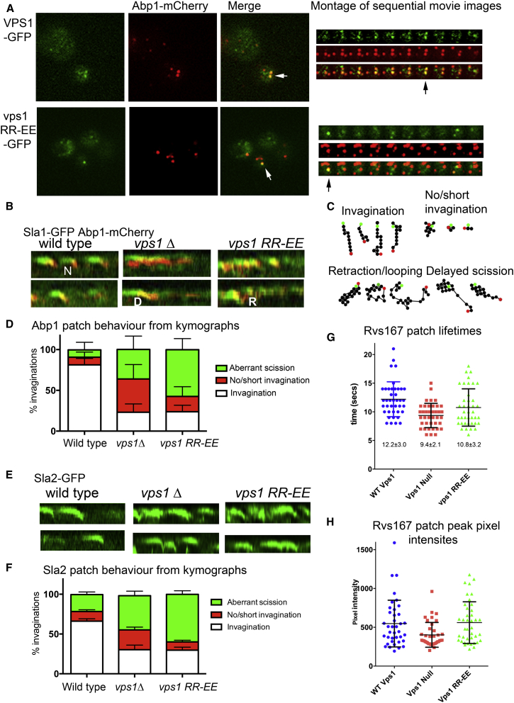
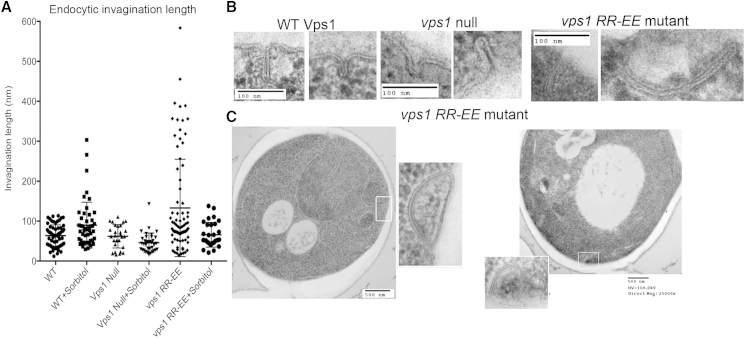
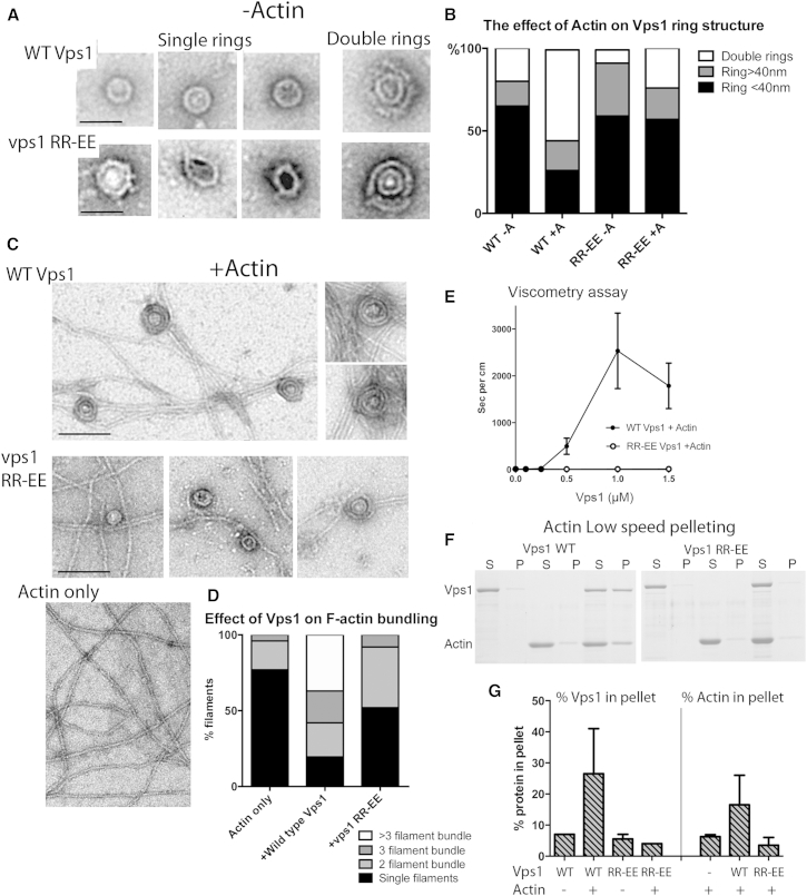
Actin is critical for endocytosis in yeast cells, and also in mammalian cells under tension. However, questions remain as to how force generated through actin polymerization is transmitted to the plasma membrane to drive invagination and scission. Here, we reveal that the yeast dynamin Vps1 binds and bundles filamentous actin. Mutational analysis of Vps1 in a helix of the stalk domain identifies a mutant RR457-458EE that binds actin more weakly. In vivo analysis of Vps1 function demonstrates that the mutation disrupts endocytosis but not other functions of Vps1 such as vacuolar trafficking or peroxisome fission. The mutant Vps1 is stably expressed in cells and co-localizes with the endocytic reporters Abp1 and the amphiphysin Rvs167. Detailed analysis of individual endocytic patch behavior indicates that the mutation causes aberrant movements in later stages of endocytosis, consistent with a scission defect. Ultrastructural analysis of yeast cells using electron microscopy reveals a significant increase in invagination depth, further supporting a role for the Vps1-actin interaction during scission. In vitro analysis of the mutant protein demonstrates that--like wild-type Vps1--it is able to form oligomeric rings, but, critically, it has lost its ability to bundle actin filaments into higher-order structures. A model is proposed in which actin filaments bind Vps1 during invagination, and this interaction is important to transduce the force of actin polymerization to the membrane to drive successful scission.


Figures

None
Graphical abstract
Figure 1
Figure 1
Wild-Type and Mutant Vps1 Interact with Actin (A) Vps1 (1.5 μM) was incubated with 3 μM yeast F-actin before high-speed centrifugation. Pellets and supernatants of samples containing Vps1 alone, actin alone, or Vps1+actin were separated by SDS-PAGE. (B) Densitometry analysis of multiple actin pelleting assays with a range of Vps1 concentrations allowed generation of a binding curve to calculate binding affinity. Error bars are SEM. (C) Crystal structure of Dynamin (PDB 3SNH) with dynamin positions denoted in black for equivalent mutagenized Vps1 residues (red). Inset reveals mutagenized residues projecting away from the stalk domain. (D) Sequence alignment of stalk region shown to bind actin in Dynamin-1. Accession numbers in sequence order: NP004399.2, NP001005336.1, NP001005360, NP001005361, Vps1 CAA82071. Vps1 residues mutated in this study are highlighted and shown with arrows. Blue represents basic residues and red represents acidic residues mutated. (E) Vps1 mutants incubated with yeast F-actin before centrifugation. Pellets and supernatants were separated by SDS-PAGE. (F) Vps1 in the pellet was analyzed using densitometry. n ≥ 3 for each mutant. Error bar is SE. Asterisks indicate level of statistical significance of differences compared to wild-type control; p value from unpaired t test 0.009 for RR-EE and 0.0001 for E473K.
Figure 2
Figure 2
The Effect of Vps1 Mutations on Functions Requiring Vps1 (A) Whole-cell extracts from yeast expressing mutated Vps1, separated by SDS-PAGE, blotted, and probed with anti-Vps1 antibodies. Apart from KRR-EEE, all mutants expressed at normal levels. Loading control is Ponceau-stained blot. (B) The effect of mutations on growth at a range of temperatures for cells expressing integrated vps1 mutants. Shown is growth at 37°C in the presence and absence of the osmotic support molecule sorbitol. (C) Vps1 is required for normal vacuolar morphology, recycling of the snare Snc1 through endosomes, and for peroxisomal fission. The effect of mutations on these functions was assessed using FM4-64, GFP-Snc1, and GFP-PTS1, respectively, and representative data are shown in upper, middle, and lower panels. (D) Carboxypeptidase Y normally functions in the vacuole where it is cleaved to its mature form (mCPY). In the absence of vps1, and in the KRR and K473E mutants, some of this material accumulates in a precursor form (pCPY). Below is a GAPDH loading control. (E) Uptake of the fluid phase marker lucifer yellow was analyzed after incubation at 21°C for up to 90 min. Localization of the majority of the dye to vacuoles (large round or lobed structure) was counted. n = 100 cells in each of two independent experiments. Errors are SD. (F) Images of cells at the 60 min time point with lucifer yellow.
Figure 3
Figure 3
The Effect of Vps1 RR-EE Mutation on Localization and Individual Endocytic Events (A) Wild-type and RR-EE Vps1 tagged with GFP on plasmids under control of the Vps1 promoter were transformed into cells also expressing the endocytic reporter Abp1 tagged with mCherry. Cells were visualized in TIRF. White arrows indicate colocalized patches. A montage from a movie for each is shown to the right. Black arrow indicates the time from which the left image was recorded. (B) Sla1-GFP (endocytic coat marker) Abp1-mCherry (actin marker) were analyzed in live cells. Shown are representative 60-s kymographs. N shows a normal invagination, R indicates example of retraction, and D delayed scission. (C) The behavior of patches was analyzed from kymographs and by generation of patch tracks. Shown are representative patch tracks for categories of Abp1 patch behaviors. Green spots indicate start of track and red spots the end. (D) Abp1 patches were categorized as showing normal invagination, no or short invagination, or normal invagination followed by aberrant scission. This latter category included retraction and delayed scission. Shown is analysis of nine or more patches in ≥17 cells from two independent experiments. Error SD. The endocytic coat protein Sla2 was also analyzed for its behavior at endocytic sites. (E) Representative 90 s kymographs from time-lapse movies of Sla2 in cells expressing wild-type or Vps1 RR-EE. (F) Graph summarizing patch behavior. Shown is analysis of ≥22 patches in seven or more cells from two independent experiments. Error is SD. (G) Lifetimes of Rvs167-GFP at endocytic sites were recorded (≥40 patches in ten cells for each strain). Shown is average lifetime ±SE. In a one-way ANOVA, lifetime is significantly different between wild-type and null (p < 0.05) but not the RR-EE mutant. (H) Peak pixel intensity of each spot was recorded as a measure of the maximum level of Rvs167 recruited to the site (±SE). One way ANOVA indicates Rvs167 intensities in the null is significantly reduced compared to cells with wild-type or vps1 RR-EE mutant (p ≥ 0.05).
Figure 4
Figure 4
The Effect of the Vps1 RR-EE Mutation on Ultrastructure of Endocytic Invaginations (A) Cells expressing wild-type or mutant Vps1 proteins were grown to log phase and processed for electron microscopy. At least 30 invaginations in at least 100 cell sections were analyzed for each strain. The effect of sorbitol on invagination length was analyzed with invagination length measured for ≥25 invaginations for each strain. All data points are shown and error bars are SD. Reduction in invagination length in the presence of sorbitol is significant in an unpaired t test (p = 0.036). (B) Examples of invaginations in wild-type and mutant cells. (C) Examples of long invaginations in the vps1 RR-EE mutant that appear to show curvature of the tubule back to the membrane.
Figure 5
Figure 5
Mutant Vps1 Is Impaired in Binding and Bundling of F-Actin (A) Vps1 was purified and visualized by EM. Shown are examples of single- and double-ring structures for wild-type and vps1 RR-EE. (B) The proportion of single and double-ring structures in the presence and absence of actin. Rings with diameter >40 nm with less regular structure were called loose rings. (C) EM images of actin alone or following incubation with wild-type or mutant Vps1. (D) In the presence of Vps1, actin was reorganized with an increased incidence of bundles. Single filaments or bundles with two, three, or more than three actin filaments were counted. (E) A falling-ball viscometry assay was performed with increasing concentrations of wild-type or mutant Vps1. Shown is a plot of the rate of fall of the ball for each concentration of Vps1. Error bars are SE; n = 3 independent experiments. (F) A low-speed actin pelleting assay with wild-type and mutant Vps1. (G) The proportion of both Vps1 (left) and actin (right) in the pellets from three independent pelleting assays was assessed using densitometry. Error is SD.

References

    1. Carroll S.Y., Stimpson H.E.M., Weinberg J., Toret C.P., Sun Y., Drubin D.G. Analysis of yeast endocytic site formation and maturation through a regulatory transition point. Mol. Biol. Cell. 2012;23:657–668. - PMC - PubMed
    1. Robertson A.S., Smythe E., Ayscough K.R. Functions of actin in endocytosis. Cell. Mol. Life Sci. 2009;66:2049–2065. - PMC - PubMed
    1. Urbanek A.N., Smith A.P., Allwood E.G., Booth W.I., Ayscough K.R. A novel actin-binding motif in Las17/WASP nucleates actin filaments independently of Arp2/3. Curr. Biol. 2013;23:196–203. - PubMed
    1. Weinberg J., Drubin D.G. Clathrin-mediated endocytosis in budding yeast. Trends Cell Biol. 2012;22:1–13. - PMC - PubMed
    1. Boulant S., Kural C., Zeeh J.C., Ubelmann F., Kirchhausen T. Actin dynamics counteract membrane tension during clathrin-mediated endocytosis. Nat. Cell Biol. 2011;13:1124–1131. - PMC - PubMed

Publication types

MeSH terms

LinkOut - more resources