

Comparative floral spur anatomy and nectar secretion in four representatives of Ranunculaceae
- PMID: 25772682
- PMCID: PMC4628095
- DOI: 10.1007/s00709-015-0794-5
Comparative floral spur anatomy and nectar secretion in four representatives of Ranunculaceae
Abstract
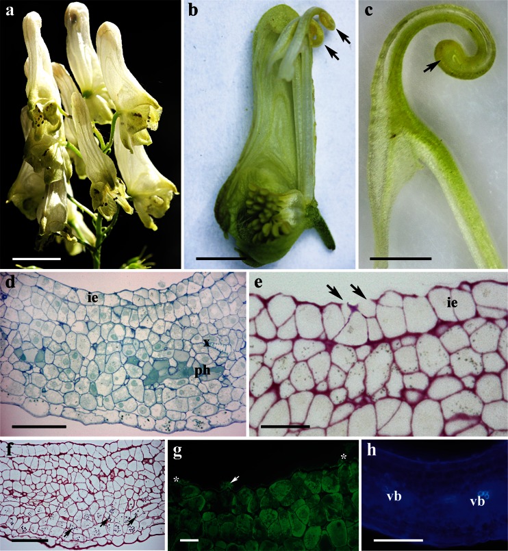
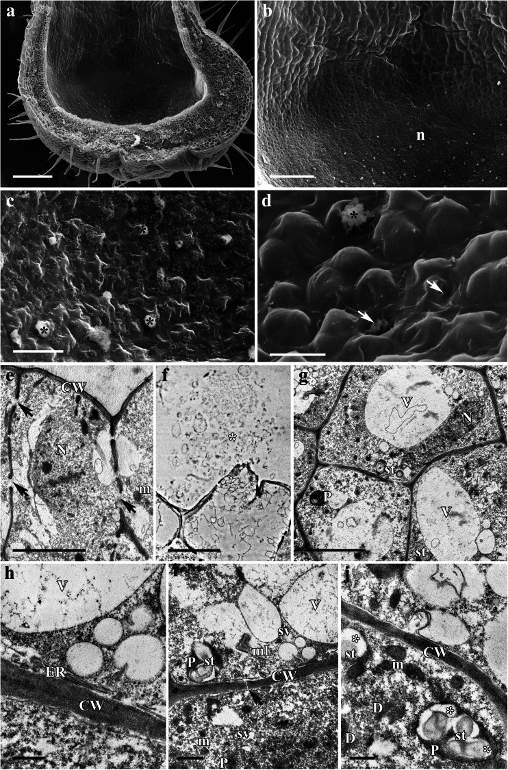
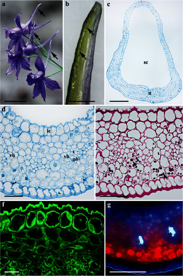
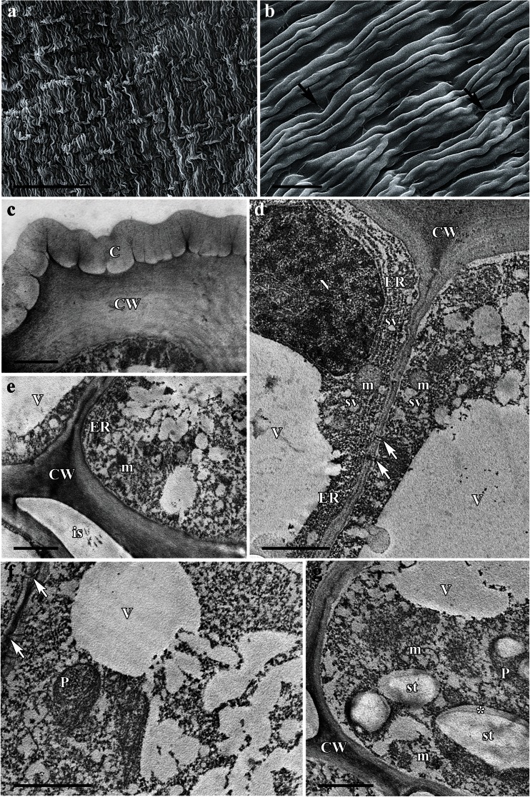
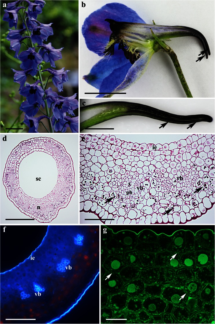
Nectaries are common in Ranunculaceae. These secretory structures, however, have not been studied in detail despite their importance in plant-animal interactions, and data relating to the structure of nectary spurs, which are so characteristic of several genera of this family, remain scarce. In order to redress this imbalance, we sought, in the present paper, to analyze the anatomical and ultrastructural organization of the nectary spurs of four representatives of Ranunculaceae, i.e., Aconitum lycoctonum L., Aquilegia vulgaris L., Consolida regalis Gray, and Delphinium elatum L. Nectary spurs were examined using light, fluorescence, scanning electron, and transmission electron microscopy. The floral nectaries of A. lycoctonum and A. vulgaris are situated at the apices of the spurs, whereas in C. regalis and D. elatum, the nectary is located along the floor surface of the spurs. Nectar in C. regalis and D. elatum is exuded through micro-channels in the cuticle, whereas in A. lycoctonum and A. vulgaris, it is released by means of cell wall disruption, indicating that the method of nectar secretion here is holocrine. Structurally, the nectary of all four investigated species is quite similar, and its cells are typical of nectar-producing cells described in the literature. It is proposed that in A. lycoctonum and A. vulgaris, disruption of the cell wall and the release of the entire cell contents into the spur cavity contribute to the composition of the nectar that the latter contains, enriching it with cytoplasmic components. We conclude that the manner of nectar exudation may vary considerably between closely related plant species, regardless of their geographical origin and phylogeny.
Keywords: Cell ultrastructure; Cuticle micro-channels; Holocrine secretion; Morphology and anatomy; Nectary structure; Secretory structures.
Figures


References
-
- Antoń S, Denisow B. Nectar production and carbohydrate composition across floral sexual phases: contrasting patterns in two protandrous Aconitum species (Delphinieae, Ranunculaceae) Flora. 2014;209:464–470. doi: 10.1016/j.flora.2014.07.001. - DOI
-
- Antoń S, Denisow B, Milaniuk K. Flowering, pollen production and insect visitation in two Aconitum species (Ranunculaceae) Acta Agrobot. 2014;67:3–12. doi: 10.5586/aa.2014.020. - DOI
-
- Bernardello G. A systematic survey of floral nectaries. In: Nicolson SW, Nepi M, Pacini E, editors. Nectaries and nectar. Dordrecht: Springer; 2007. pp. 19–128.
-
- Canto A, Herrera CM, García IM, Pérez R, Vaz M. Intraplant variation in nectar traits in Helleborus foetidus (Ranunculaceae) as related to floral phase, environmental conditions and pollinator exposure. Flora. 2011;206:668–675. doi: 10.1016/j.flora.2011.02.003. - DOI
-
- Considine JA, Knox RB. Development and histochemistry of the cells, cell walls, and cuticle of the dermal system of fruit of the grape Vitis vinifera L. Protoplasma. 1979;99:347–365. doi: 10.1007/BF01275807. - DOI
Publication types
MeSH terms
Substances
LinkOut - more resources
Full Text Sources
Other Literature Sources
