. 2015 May;129(5):653-67.
doi: 10.1007/s00401-015-1404-5. Epub 2015 Mar 15.

M. leprae components induce nerve damage by complement activation: identification of lipoarabinomannan as the dominant complement activator

Affiliations

M. leprae components induce nerve damage by complement activation: identification of lipoarabinomannan as the dominant complement activator

Nawal Bahia El Idrissi et al. Acta Neuropathol. 2015 May.

Abstract

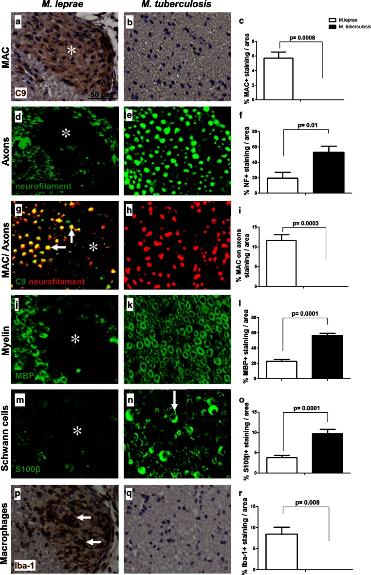
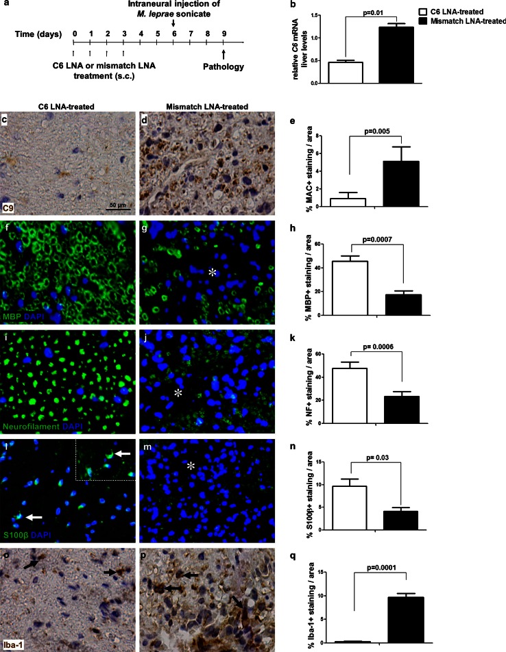
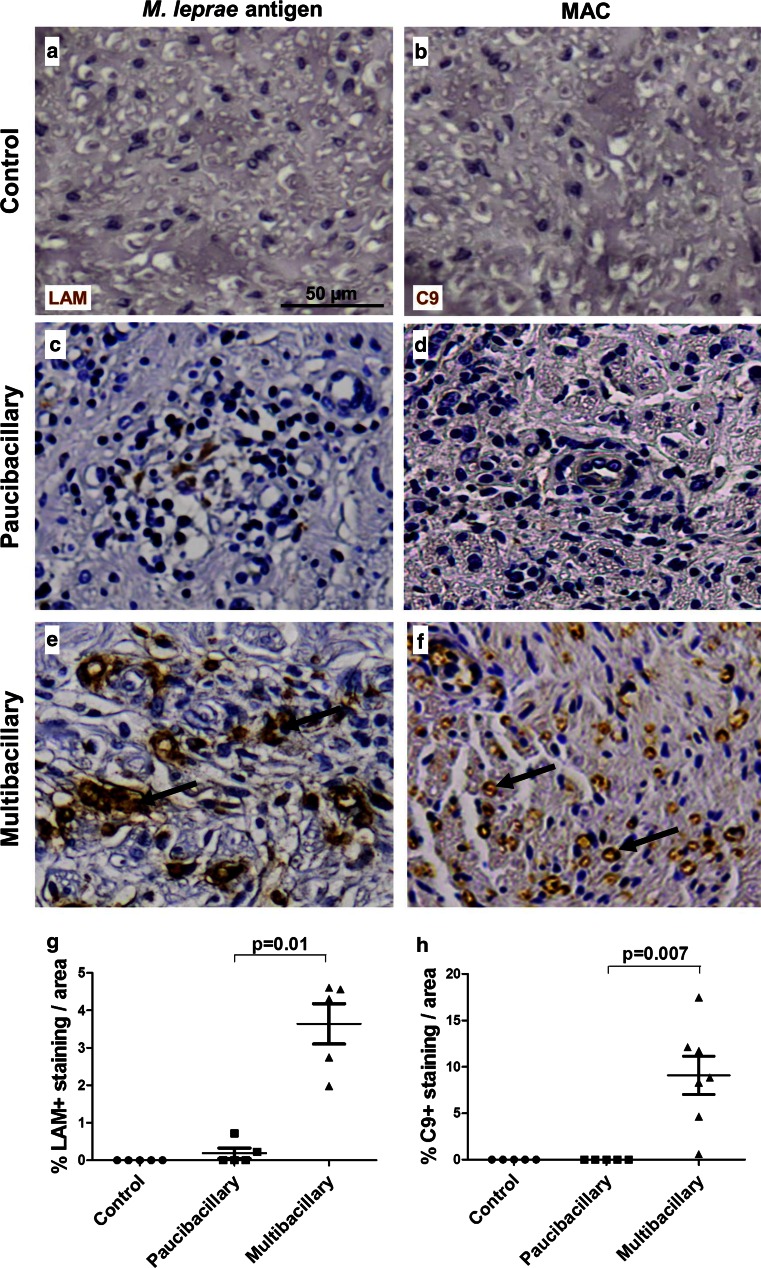
Peripheral nerve damage is the hallmark of leprosy pathology but its etiology is unclear. We previously identified the membrane attack complex (MAC) of the complement system as a key determinant of post-traumatic nerve damage and demonstrated that its inhibition is neuroprotective. Here, we determined the contribution of the MAC to nerve damage caused by Mycobacterium leprae and its components in mouse. Furthermore, we studied the association between MAC and the key M. leprae component lipoarabinomannan (LAM) in nerve biopsies of leprosy patients. Intraneural injections of M. leprae sonicate induced MAC deposition and pathological changes in the mouse nerve, whereas MAC inhibition preserved myelin and axons. Complement activation occurred mainly via the lectin pathway and the principal activator was LAM. In leprosy nerves, the extent of LAM and MAC immunoreactivity was robust and significantly higher in multibacillary compared to paucibacillary donors (p = 0.01 and p = 0.001, respectively), with a highly significant association between LAM and MAC in the diseased samples (r = 0.9601, p = 0.0001). Further, MAC co-localized with LAM on axons, pointing to a role for this M. leprae antigen in complement activation and nerve damage in leprosy. Our findings demonstrate that MAC contributes to nerve damage in a model of M. leprae-induced nerve injury and its inhibition is neuroprotective. In addition, our data identified LAM as the key pathogen associated molecule that activates complement and causes nerve damage. Taken together our data imply an important role of complement in nerve damage in leprosy and may inform the development of novel therapeutics for patients.


Figures

Fig. 1
Fig. 1
M. leprae induces complement deposition and nerve damage in vivo. Immunohistochemistry and quantification for C9 detecting MAC (ac), neurofilament detecting axons (cf), co-localization of MAC and axons (gi), MBP detecting myelin (jl), S100β detecting Schwann cells (mo), or Iba-1 detecting macrophages (pr) in cross sections of mouse sciatic nerves at 3 days post-injection with either M. leprae (a, d, g, j, m, p) or M. tuberculosis (b, e, h, k, n, q), showing a significant higher amount of MAC immunoreactivity (a, asterisk) (c, Student’s t test: p = 0.0008), axonal damage (d) and loss (d, asterisk) (f, Student’s t test: p = 0.01), MAC deposited on axons (g, arrows) (i, Student’s t test: p = 0.0003) and axonal debris (g, asterisk), myelin loss (j, asterisk) (l, Student’s t test: p = 0.0001), loss of S100β expression on Schwann cells (m, asterisk) (o, Student’s t test: p = 0.0001) and accumulation of macrophages (p, arrows) (r, Student’s t test: p = 0.008) in M. leprae-injected nerves compared to M. tuberculosis-injected nerves where no MAC deposition and nerve damage was detected (b, e, h, k, n, q). The arrow in (n) points to the normal moon-shaped appearance of S100β-positive Schwann cells
Fig. 2
Fig. 2
The M. leprae component lipoarabinomannan (LAM) is the dominant complement activator and induces nerve damage in vivo. a Haemolytic assay of normal human serum (NHS) pre-incubated for 1 h at 37 °C with either M. leprae sonicate (5 µg/µl) or M. tuberculosis sonicate (5 µg/µl) or PBS as controls, showing significantly decreased haemolytic activity in NHS pre-incubated with M. leprae but not with M. tuberculosis or PBS, demonstrating complement consumption by M. leprae. b ELISA for MAC generation on M. leprae sonicate (2.5 µg)-coated plates incubated with either mannose binding lectin deficient (MBL−/−) serum (to test for the contribution of the lectin pathway) or NHS in the presence of the neutralizing anti-C1q antibody (to test for the contribution of the classical pathway) or C1 inhibitor (to test for the combined contribution of the lectin and classical pathways) or BSA as control, showing a significant reduction of MAC formation in the MBL−/− serum and NHS supplemented with the C1 inhibitor, but not by the neutralizing anti-C1q antibody, demonstrating complement activation by M. leprae via the lectin pathway. c ELISA for TCC generation in NHS on plates coated with either M. leprae sonicate (2.5 µg) or its cellular fractions, including cell membrane (2.5 µg), the inner cell wall component lipoarabinomannan (LAM) (2.5 µg) or the outer cell wall component phenolic glycolipid 1 (PGL-1) (2.5 µg), showing that all components except PGL-1 result in TCC generation. dr Intraneural injections of cell membrane or LAM induce complement deposition and nerve damage in vivo. Immunohistochemistry and quantification for C9 detecting MAC (dg), neurofilament detecting axons (hk), MBP detecting myelin (lo), S100β detecting Schwann cells (ps) or Iba-1 detecting macrophages (tw) in cross sections of mouse sciatic nerves at 72 h post-injection with either PBS (d, h, l, p, t), cell membrane (e, i, m, q, u) or LAM (f, j, n, r, v), showing a significant higher amount of MAC deposition (e, f, asterisks) (g, One way ANOVA test: p = 0.0001; p = 0.0001), axonal damage (i, j, asterisks) (k, One way ANOVA test: p = 0.0001; p = 0.0001), loss of myelin proteins (m, n, asterisks) (o, One way ANOVA test: p = 0.0001; p = 0.0001), loss of S100β expression on Schwann cells (q, r, asterisks) (s, One way ANOVA test: p = 0.0001; p = 0.0001) and accumulation of macrophages (u, v, arrows) in cell membrane- and LAM-injected nerves compared to PBS-injected nerves where no signs of MAC deposition (d), undamaged nerve morphology (h, l), preserved S100β expression (p) and a paucity of endoneurial macrophages (t) were observed
Fig. 3
Fig. 3
MAC inhibition by C6 antisense therapy protects against M. leprae-induced nerve damage. a Schedule of treatment and experimental timeline for the C6 antisense therapy. Mice were treated for 4 days with either the C6 LNA (n = 5) or the control mismatch LNA (n = 5). At day 6, M. leprae sonicate was injected into the mouse sciatic nerve. At day 9 (3 days post-injection) mice were sacrificed for determination of C6 mRNA liver levels and pathological analysis. b qPCR of liver C6 mRNA, showing significant lower levels in mice treated with the C6 LNA compared to mismatch LNA-treated controls. Immunohistochemistry and quantification of C9 detecting MAC (ce), MBP detecting myelin (fh), neurofilament detecting axons (ik), S100β detecting Schwann cells (ln) or Iba-1 detecting macrophages (oq) in cross sections of sciatic nerves from C6 LNA-treated (c, f, i, l, o) or mismatch LNA-treated (d, g, j, m, p) mice at 72 h post-injection with M. leprae sonicate, showing a significant and robust reduction in MAC deposition (Student’s t test: p = 0.005) (e), intact myelin (Student’s t test: p = 0.0007) (h) and axonal morphology (k), S100β expression by Schwann cells (l, arrows) and reduced accumulation of macrophages (Student’s t test: p = 0.0001) (k) in C6 LNA-treated mice compared to mismatch-treated controls (asterisks in g, j, m indicate damaged areas of the mismatch-treated nerves, arrows in p indicate iba-1 positive macrophages in the mismatch-treated nerves)
Fig. 4
Fig. 4
LAM and MAC deposition in nerves of leprosy patients. Immunostaining for the M. leprae antigen LAM and C9, detecting MAC, in nerve biopsies of control (a, b) compared to paucibacillary (c, d) and multibacillary (e, f) leprosy patients. The control nerves were negative for LAM (a) and MAC (b), as expected. Paucibacillary nerves show little immunoreactivity for LAM (c) and virtually no MAC deposition (d), whereas multibacillary patients show robust staining for LAM (e, arrows) and MAC (f). Quantification of the immunostaining showed that the amount of immunoreactivity for LAM (g) and MAC (h) is significantly higher in multibacillary compared to paucibacillary nerves (Student’s t test paucibacillary vs. multibacillary: LAM, p = 0.01; C9, p = 0.007). Error bars indicate standard error of the mean
Fig. 5
Fig. 5
LAM is associated with MAC deposition in nerves of leprosy patients. Immunofluorescent double staining for complement component C9, detecting MAC, and the M. leprae antigen LAM, showing co-localization in the nerves of multibacillary patients (a). C9 and LAM also co-localized with the SMI31 (b) and the neurofilament (NF) (c) markers of axons, respectively. The amount of C9 immunoreactivity significantly correlated with the amount of LAM immunoreactivity found in paucibacillary and multibacillary leprosy nerves (Pearson’s correlation, r = 0.9601, p < 0.0001) (d), indicating an association between the extent of M. leprae antigen LAM and MAC deposition in leprosy nerves

References

    1. Ridley DS, Jopling WH. Classification of leprosy according to immunity. A five-group system. Int J Lepr Other Mycobact Dis. 1966;34:255–273. - PubMed
    1. Laal S, Bhutani LK, Nath I. Natural emergence of antigen-reactive T cells in lepromatous leprosy patients during erythema nodosum leprosum. Infect Immun. 1985;50:887–892. - PMC - PubMed
    1. Sohi AS, Kandhari KC, Singh N. Motor nerve conduction studies in leprosy. Int J Dermatol. 1971;10:151–155. doi: 10.1111/j.1365-4362.1971.tb03727.x. - DOI - PubMed
    1. Rambukkana A, Zanazzi G, Tapinos N, Salzer JL. Contact-dependent demyelination by Mycobacterium leprae in the absence of immune cells. Science. 2002;296:927–931. doi: 10.1126/science.1067631. - DOI - PubMed
    1. Shetty VP, Mistry NF, Birdi TJ, Antia NH. Effect of T-cell depletion on bacterial multiplication and pattern of nerve damage in M. leprae-infected mice. Indian J Lepr. 1995;67:363–374. - PubMed

Publication types

MeSH terms

Substances