

In vivo cell tracking with bioluminescence imaging
- PMID: 25774232
- PMCID: PMC4354780
- DOI: 10.1007/s13139-014-0309-x
In vivo cell tracking with bioluminescence imaging
Abstract
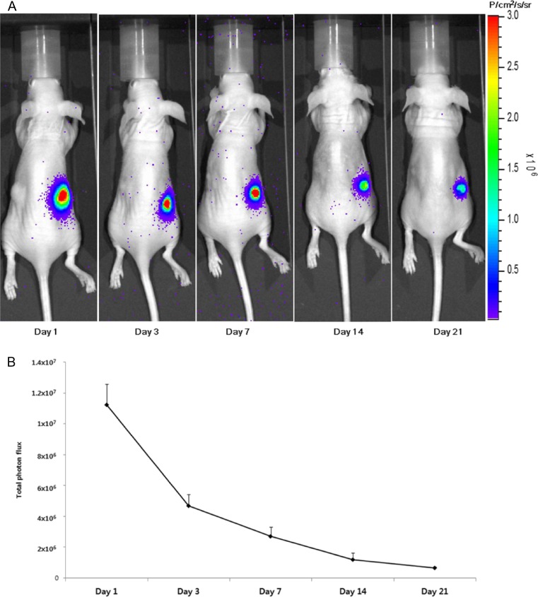
Molecular imaging is a fast growing biomedical research that allows the visual representation, characterization and quantification of biological processes at the cellular and subcellular levels within intact living organisms. In vivo tracking of cells is an indispensable technology for development and optimization of cell therapy for replacement or renewal of damaged or diseased tissue using transplanted cells, often autologous cells. With outstanding advantages of bioluminescence imaging, the imaging approach is most commonly applied for in vivo monitoring of transplanted stem cells or immune cells in order to assess viability of administered cells with therapeutic efficacy in preclinical small animal models. In this review, a general overview of bioluminescence is provided and recent updates of in vivo cell tracking using the bioluminescence signal are discussed.
Keywords: Bioluminescence; Cell therapy; Cell tracking; In vivo imaging; Optical imaging.
Figures

References
-
- Berman SC, Galpoththawela C, Gilad AA, Bulte JW, Walczak P. Long-term MR cell tracking of neural stem cells grafted in immunocompetent versus immunodeficient mice reveals distinct differences in contrast between live and dead cells. Magn Reson Med. 2011;65(2):564–74. doi: 10.1002/mrm.22613. - DOI - PMC - PubMed
-
- Acton P, Zhou R. Imaging reporter genes for cell tracking with PET and SPECT. Q J Nucl Med Mol Imaging. 2005;49(4):349–60. - PubMed
Publication types
LinkOut - more resources
Full Text Sources
Other Literature Sources