

Case Reports
doi: 10.1007/s13139-014-0295-z.
Epub 2014 Sep 25.
Usefulness of (131)I-SPECT/CT and (18)F-FDG PET/CT in Evaluating Successful (131)I and Retinoic Acid Combined Therapy in a Patient with Metastatic Struma Ovarii
Affiliations
- PMID: 25774238
- PMCID: PMC4354789
- DOI: 10.1007/s13139-014-0295-z
Item in Clipboard
Case Reports
Usefulness of (131)I-SPECT/CT and (18)F-FDG PET/CT in Evaluating Successful (131)I and Retinoic Acid Combined Therapy in a Patient with Metastatic Struma Ovarii
Nucl Med Mol Imaging.
2015 Mar.
Abstract
Metastatic struma ovarii is an extremely rare disease, and the treatment of choice has not been established. Here, we introduce the case of a 36-year-old female pregnant patient with metastatic struma ovarii. Initial treatment was an exploratory laparotomy to remove multiple peritoneal masses. After delivery, a total thyroidectomy was done for the further (131)I-therapy. (131)I-SPECT/CT and (18) F-FDG PET/CT showed multiple hepatic metastases and extensive peritoneal seeding nodules. Multiple (131)I and retinoic acid combination therapies were performed, resulting in marked improvement. (131)I-SPECT/CT and (18) F-FDG PET/CT were quite useful for evaluating the biologic characteristics of the metastases.
Figures
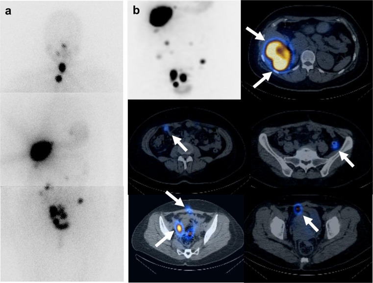
The first post-therapy 131I whole-body scan and 131I-SPECT/CT. a Post-therapy 131I whole-body images (30 mCi) show remnant thyroid glands. Intense iodine-avid lesions in the right lobe of the liver and multiple peritoneal seedings are well visualized. b Post-therapy 131I-SPECT/CT of the abdominopelvic cavity was obtained. The maximized intensity projection image is located in the left upper row. More prominent uptake in the multiple hepatic and peritoneal seedings is shown because of the longer acquisition time and three-dimensional modality. On the fusion axial image of 131I-SPECT/CT, hepatic metastases show iodine-avid uptake. Second and third row images show multiple peritoneal seedings with avid iodine uptake regardless of the small size (white arrow)
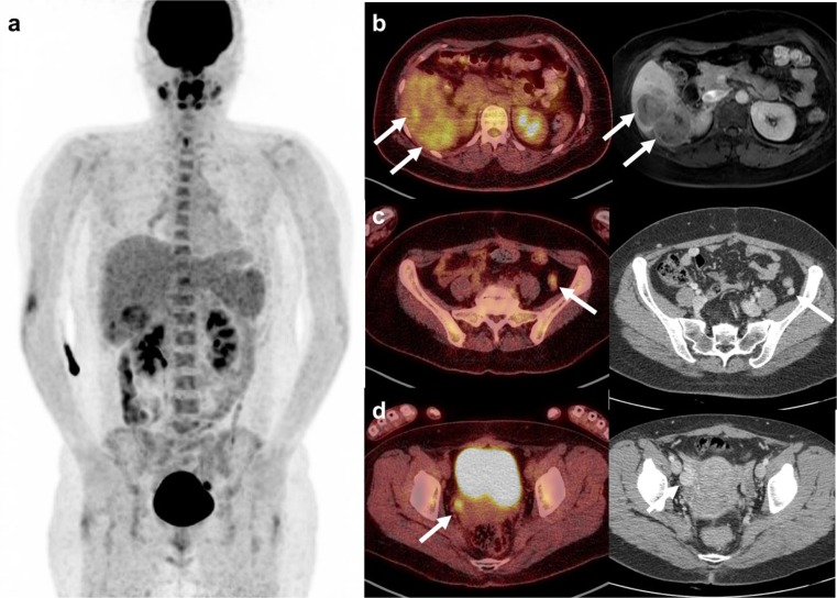
18F-FDG PET/CT after the first 131I therapy. a Maximized intensity projection image shows hepatic metastasis in the right lobe of the liver and several seeding nodules with variable FDG uptake. b Fusion axial image shows a different glucose metabolism. The S5 lesion shows mild FDG uptake, but the S6 lesion shows intense FDG uptake. Multiple heptatic metastases were well correlated with MRI. c A small peritoneal seeding in the left iliac fossa shows increased uptake on the fusion PET/CT image and good enhancement on contrast-enhanced CT. d Intense uptake in the peritoneal seeding in the right pelvic side wall is visualized on the fusion PET/CT image. A round lesion with good enhancement is noted on contrast-enhanced CT
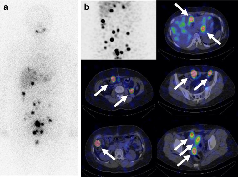
The second post-therapy 131I whole-body scan and 131I-SPECT/CT combined with retinoic acid. a After the removal of hepatic metastases, post- 131I therapy a whole-body scan (200 mCi) was performed. More prominent uptake in the multiple peritoneal seedings is visualized in the perihepatic and abdominopelvic cavity. b
131I-SPECT/CT shows intense iodine uptake in the multiple peritoneal seeding nodules. A maximized intensity projection image of the abdominopelvic cavity is demonstrated in the left upper row. Fusion axial images of 131I-SPECT/CT reveal the location of peritoneal seedings in the perihepatic area, perigastric area, right and lower quadrant pelvic cavity, rectovaginal pouch, etc.
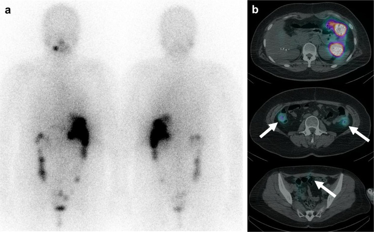
The fifth post-therapy 131I whole-body scan and 131I-SPECT/CT. a Post-therapy 131I whole-body scan (200 mCi) reveals physiologic stomach and bowel uptake. Suspicious uptake is visualized in the pelvic cavity. b Fusion axial images of 131I-SPECT/CT show matched physiologic bowel uptake in the upper row and faint uptake in the previous seeding lesions in the pelvic cavity in the middle and lower row (white arrow). There was no significant lesion with avid iodine uptake. The patient showed a markedly improved state
References
-
- Navarro MD, Tan MA, Lovecchio JL, Hajdu SI. Case report: malignant struma ovarii. Ann Clin Lab Sci. 2004;34(1):107–112. - PubMed
Publication types
LinkOut - more resources
Full Text Sources
Other Literature Sources