

Pleura space anatomy
- PMID: 25774304
- PMCID: PMC4332049
- DOI: 10.3978/j.issn.2072-1439.2015.01.48
Pleura space anatomy
Abstract
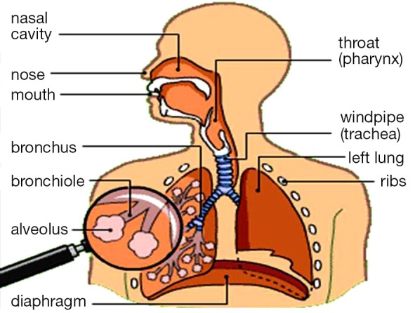
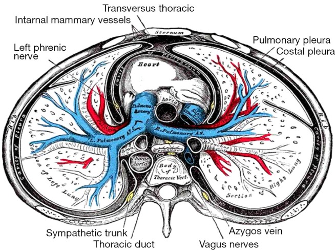
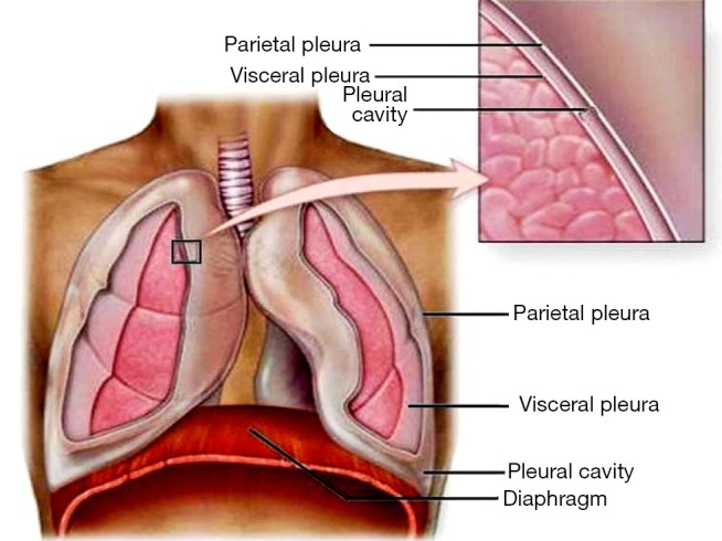
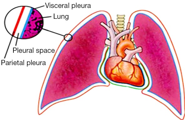
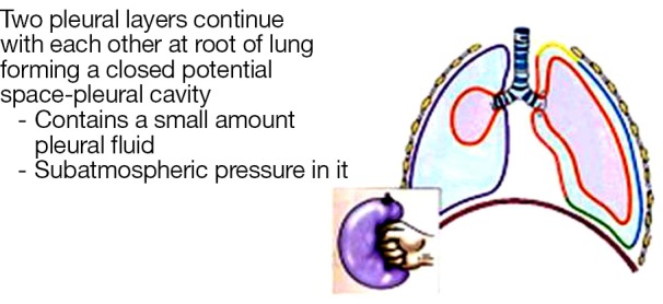
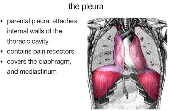
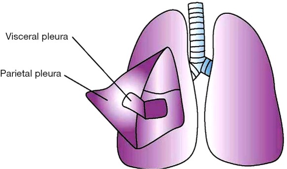
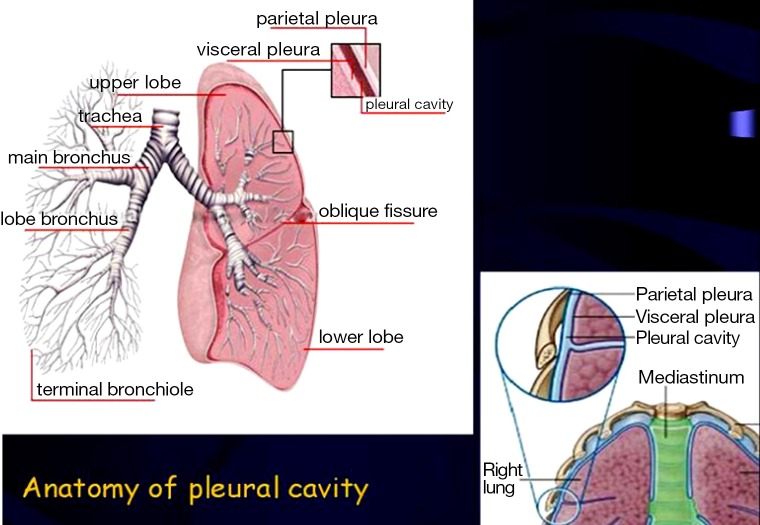
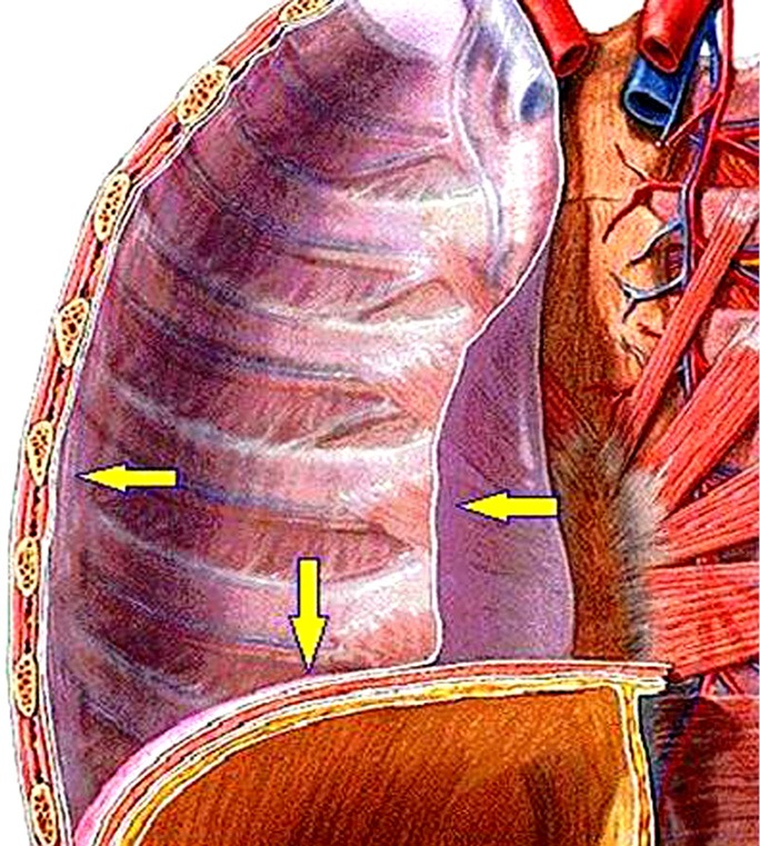
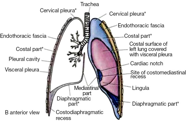
The pleural cavity is the potential space between the two pleurae (visceral and parietal) of the lungs. The pleurae are serous membranes which fold back onto themselves to form a two-layered membranous structure. The thin space between the two pleural layers is known as the pleural cavity and normally contains a small amount of pleural fluid. There are two layers; the outer pleura (parietal pleura) is attached to the chest wall and the inner pleura (visceral pleura) covers the lungs and adjoining structures, via blood vessels, bronchi and nerves. The parietal pleurae are highly sensitive to pain, while the visceral pleura are not, due to its lack of sensory innervation. In the current review we will present the anatomy of the pleural space.
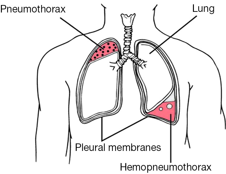
Keywords: Pneumothorax; anatomy; pleural space.
Figures

References
-
- Williams PL, Warwick R, Dyson M, et al. eds. Gray’s Anatomy. Edinburgh: Churchill Livingstone, 1995.
-
- Murray IF, Nadel IA. eds. Textbook of Respiratory Medicine. Philadelphia: Saunders, 1988.
-
- Wang NS. Anatomy and physiology of the pleural space. Clin Chest Med 1985;6:3-16. - PubMed
-
- Lee KF, Olak J. Anatomy and physiology of the pleural space. Chest Surg Clin N Am 1994;4:391-403. - PubMed
Publication types
LinkOut - more resources
Full Text Sources
Other Literature Sources