

Physiology of the pleural space
- PMID: 25774305
- PMCID: PMC4332077
- DOI: 10.3978/j.issn.2072-1439.2014.12.48
Physiology of the pleural space
Abstract
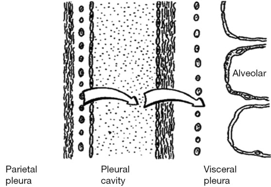
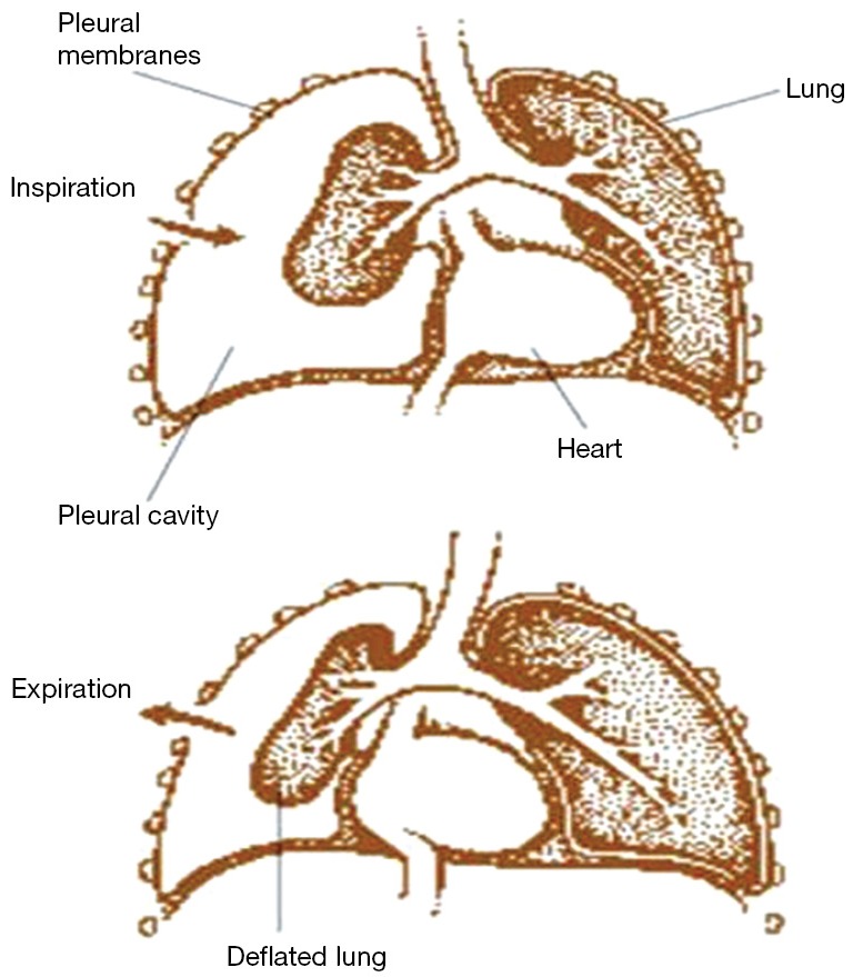
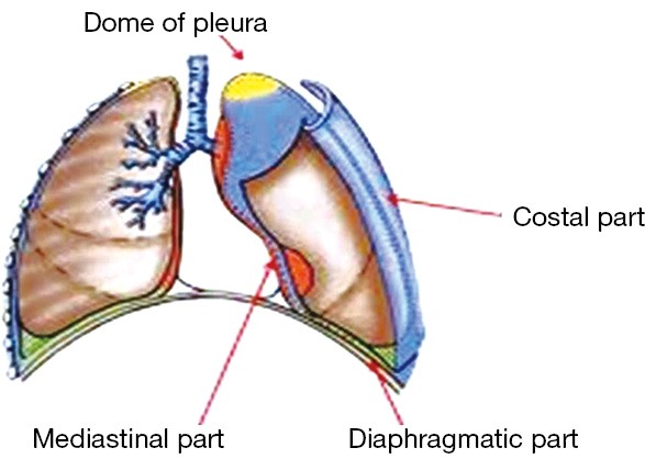
The pleural cavity is created between the 4(th) and 7(th) week of embryologic development. These embryonic components of visceral and parietal pleurae develop different anatomic characteristics with regard to vascular, lymphatic, and nervous supply. There are two layers: a superficial mesothelial cell layer facing the pleural space and an underlying connective tissue layer. The pleura might present inflammatory response and maintenance of the pleural fluid is observed. The latter function is especially important in the mechanical coupling of the lung and chest wall. Fluid is filtered into the pleural space according to the net hydrostatic oncotic pressure gradient. It flows downward along a vertical pressure gradient, presumably determined by hydrostatic pressure and resistance to viscous flow. There also may be a net movement of fluid from the costal pleura to the mediastinal and interlobar regions. In these areas, pleural fluid is resorbed primarily through lymphatic stomata on the parietal pleural surface. In the current review we will present the physiology of the pleural space in a step by step manner.
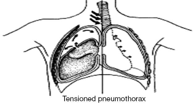
Keywords: Pneumothorax; physiology; pleural space.
Figures
References
-
- Anderson RH, Sarwark AE, Spicer DE, et al. Exercises in anatomy: holes between the ventricles. Multimed Man Cardiothorac Surg 2014;2014. - PubMed
-
- Fitting JW. From Breathing to Respiration. Respiration 2014. [Epub ahead of print]. - PubMed
-
- Wang NS. Anatomy and physiology of the pleural space. Clin Chest Med 1985;6:3-16. - PubMed
-
- Lee KF, Olak J. Anatomy and physiology of the pleural space. Chest Surg Clin N Am 1994;4:391-403. - PubMed
Publication types
LinkOut - more resources
Full Text Sources
Medical