. 2015 Apr;125(4):1569-78.
doi: 10.1172/JCI80115. Epub 2015 Mar 16.

Severe myopathy in mice lacking the MEF2/SRF-dependent gene leiomodin-3

Severe myopathy in mice lacking the MEF2/SRF-dependent gene leiomodin-3

Bercin K Cenik et al. J Clin Invest. 2015 Apr.

Abstract

Maintenance of skeletal muscle structure and function requires a precise stoichiometry of sarcomeric proteins for proper assembly of the contractile apparatus. Absence of components of the sarcomeric thin filaments causes nemaline myopathy, a lethal congenital muscle disorder associated with aberrant myofiber structure and contractility. Previously, we reported that deficiency of the kelch-like family member 40 (KLHL40) in mice results in nemaline myopathy and destabilization of leiomodin-3 (LMOD3). LMOD3 belongs to a family of tropomodulin-related proteins that promote actin nucleation. Here, we show that deficiency of LMOD3 in mice causes nemaline myopathy. In skeletal muscle, transcription of Lmod3 was controlled by the transcription factors SRF and MEF2. Myocardin-related transcription factors (MRTFs), which function as SRF coactivators, serve as sensors of actin polymerization and are sequestered in the cytoplasm by actin monomers. Conversely, conditions that favor actin polymerization de-repress MRTFs and activate SRF-dependent genes. We demonstrated that the actin nucleator LMOD3, together with its stabilizing partner KLHL40, enhances MRTF-SRF activity. In turn, SRF cooperated with MEF2 to sustain the expression of LMOD3 and other components of the contractile apparatus, thereby establishing a regulatory circuit to maintain skeletal muscle function. These findings provide insight into the molecular basis of the sarcomere assembly and muscle dysfunction associated with nemaline myopathy.


Figures

Figure 7
Figure 7. A model for the role of LMOD3 and KLHL40 in actin cycling and MRTF/SRF- and MEF2-dependent transcription.
In normal muscle cells, MRTF/SRF and MEF2 regulate LMOD3 expression, and MEF2 regulates KLHL40 expression. KLHL40 functions in the cytoplasm to stabilize LMOD3 protein. Together, they promote actin polymerization by converting G-actin to F-actin, allowing normal sarcomeric function. In muscles lacking LMOD3, accumulation of G-actin monomers not only disrupt sarcomeric integrity, but also repress MRTF-A expression, which in turn suppresses SRF-dependent target genes encoding cytoskeletal proteins and components of the contractile apparatus, leading to nemaline myopathy.
Figure 6
Figure 6. LMOD3 and KLHL40 regulation of MRTF/SRF and MEF2 pathways.
Synthetic luciferase reporters with multimerized (A) CArG boxes (SM22-4x-CArG) and (B) MEF2 sites (3x-MEF2-Luc) were transfected into COS-7 cells, together with LMOD3 and/or KLHL40 expression vectors. Cells were cotransfected with MRTF-A (A) or MEF2C (B). Experiments were performed at least twice, with all transfections done in triplicate. All results are reported as fold change ± SEM. (C and D) qPCR analysis was performed on gastrocnemius muscles from WT and KO mice for (C) Mrtf gene expression and (D) SRF target gene expression. Experiments were performed in triplicate, and expression was normalized to 18S rRNA. *P < 0.05; **P < 0.01; ***P < 0.001; ****P < 0.0001.
Figure 5
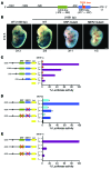
Figure 5. Regulation of Lmod3 promoter by MEF2 and SRF transcription factors.
(A) The Lmod3 promoter region contains consensus sites for MEF2 and SRF (CArG box). The MEF2 site is also a TATA box. Numbering of nucleotide positions is relative to the ATG (+1) start codon. (B) LacZ staining was performed on E12.5 F0 embryos containing the 1691- or 1069-bp enhancer fragment of the Lmod3 promoter cloned into an hsp68-LacZ construct. WT Lmod3 Tg embryos showed strong muscle-specific expression that was diminished in the SRF/MEF2 Tg mutants. Scale bar: 1 mm. (C) COS-7 cells were transfected with the upstream fragments of the Lmod3 gene cloned into the luciferase reporter plasmid pGL3. Luciferase reporter assays showed activation of the Lmod3 promoter by SRF in response to its coactivator MRTF-A. (D) Luciferase activity was measured in COS-7 cells transfected with the 1691-bp Lmod3 reporter plasmid with or without mutations in the MEF2 site and/or the CArG box. Cells were cotransfected with MEF2C. (E) Luciferase reporter activity was measured in COS-7 cells transfected with the 1691-bp Lmod3 reporter plasmid with or without mutations in the CArG box and/or the MEF2 site. Cells were cotransfected with MRTF-A. Experiments were performed at least twice, with all transfections done in triplicate. All results are reported as percentage values ± SEM (WT construct with MEF2C and MRTF-A was set to 100%). Luc, luciferase.
Figure 4
Figure 4. Loss of LMOD3 causes nemaline myopathy in mice.
(A) H&E staining of quadriceps, gastrocnemius, plantaris, and soleus (GPS) muscles from 1-, 3- and 8-week-old WT and KO mice. By 3 weeks of age, centralization of nuclei and loss of sarcomeric organization were observed in the KO mice. Scale bars: 40 μm. (B) Desmin (red), laminin (green), and DAPI (blue) immunostaining of transverse myofibers shows centralized nuclei and pathological desmin accumulation in the KO muscle. Scale bars: 40 μm. (C) Transmission electron microscopic images of longitudinal sections of the soleus muscle from 3-week-old WT and KO mice showing loss of sarcomeric organization, increased glycogen accumulation, and replacement of sarcomeric Z lines by electron-dense nemaline bodies. Dotted lines indicate healthy sarcomere in WT muscle and demarcate disorganized sarcomeres in KO muscle. White arrowheads denote Z lines in WT muscle and mark the nemaline bodies (second panel) or Z-line streaming (third and fourth panels) in KO muscle. Black arrowhead denotes glycogen granules. All histologic analyses were performed in age- and sex-matched littermates (n = 3/group). Scale bars: 1 μm.
Figure 3
Figure 3. Lmod3-KO mice have a failure-to-thrive phenotype, which is rescued by the MCK-Lmod3 transgene.
(A) Photos of 2- and 3-week-old Lmod3-KO mice with sex-matched WT littermates show significant size differences. (B) Growth curve of KO male mice and WT male littermates shows failure to thrive in the KO mice. (C) A significant difference in muscle mass was observed in the KO mice compared with that of their WT littermates (n ≥10 animals per group). **P < 0.01; *P < 0.05. MM/TL, muscle mass/tibia length. (D) Wheat-germ agglutinin (WGA) staining of muscle membranes in quadriceps of P12 mice showed reduced myofiber size in KO mice compared with that in WT littermates. WGA (red), DAPI (blue). Scale bars: 20 μm. (E) Quantification of myofiber area in quadriceps and gastrocnemius (Gastroc) muscles from transverse sections stained with laminin (shown in Figure 4B). Fiber size was calculated by particle analysis using ImageJ software. ****P < 0.0001. (F) Representative image of MCK-Lmod3 Δ2-KO mice (KO Tg) and Δ2-KO mice, together with their MCK-Lmod3 (WT Tg) and WT littermates showing complete rescue of the KO phenotype, with muscle-specific overexpression of LMOD3 with the MCK-Lmod3 transgene. (G) Body mass of KO Tg mice was indistinguishable from the body mass of WT and WT Tg mice. WT, n = 3; WT Tg, n = 3; KO, n = 3; and KO Tg, n = 3. Animals were 3-week-old males. Data are presented as the mean ± SEM. (H) Lmod3 expression in the skeletal muscle (gastrocnemius) of 3-week-old mice was assayed by qPCR. Experiments were performed in triplicate with 3 biological replicates, and expression was normalized to 18S rRNA.
Figure 2
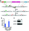
Figure 2. TALEN-induced frameshift mutagenesis eliminates LMOD3 at the RNA and protein levels.
(A) LMOD3 protein structure, intron-exon structure of the Lmod3 gene, and gene-targeting strategy. TALEN pairs targeting the second exon induce double-stranded breaks (DSB), followed by error-prone nonhomologous end-joining (NHEJ) repair. Frameshift deletions lead to premature termination of transcription. LRR, leucine-rich repeat; Tm-h, Tm-binding helix; A-h, actin-binding helix; WH2, Wiskott-Aldrich syndrome protein homology domain 2. (B) Loss of Lmod3 mRNA was confirmed in the heart and quadriceps of KO mice and in those of their control WT littermates (n = 3/group). Experiments were performed in triplicate, and expression was normalized to 18S rRNA. (C) Western blot analysis of skeletal muscle with an antibody recognizing the N-terminal of LMOD3 protein showed a complete loss of LMOD3 in KO mice. GAPDH was used as a loading control.
Figure 1
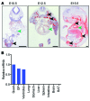
Figure 1. Lmod3 is expressed selectively in the skeletal muscle and heart.
(A) ISH analysis was performed on transverse sections of E10.5 and E12.5 embryos and sagittal sections of E15.5 embryos using radioisotopic antisense RNA probes against Lmod3. Black arrowheads denote developing muscle in E10.5 and E12.5 sections and intercostal muscles, diaphragm, and developing muscle groups in the limb bud in the E15.5 section. Green arrowheads indicate the heart. Scale bars: 500 μm. (B) qPCR analysis of Lmod3 in an adult mouse shows heart- and muscle-specific expression. Experiments were performed in triplicate, and expression was normalized to 18S rRNA. Quad, quadriceps; GP, gastrocnemius and plantaris; BAT, brown adipose tissue.

References

    1. Ryan MM, et al. Clinical course correlates poorly with muscle pathology in nemaline myopathy. Neurology. 2003;60(4):665–673. doi: 10.1212/01.WNL.0000046585.81304.BC. - DOI - PubMed
    1. Romero NB, Clarke NF. Congenital myopathies. Handb Clin Neurol. 2013;113:1321–1336. doi: 10.1016/B978-0-444-59565-2.00004-6. - DOI - PubMed
    1. Romero NB, Sandaradura SA, Clarke NF. Recent advances in nemaline myopathy. Curr Opin Neurol. 2013;26(5):519–526. doi: 10.1097/WCO.0b013e328364d681. - DOI - PubMed
    1. North KN, Ryan MM. Nemaline Myopathy. GeneReviews. Seattle, Washington, USA: University of Washington; 2002. - PubMed
    1. Nowak KJ, Ravenscroft G, Laing NG. Skeletal muscle alpha-actin diseases (actinopathies): pathology and mechanisms. Acta Neuropathol. 2013;125(1):19–32. doi: 10.1007/s00401-012-1019-z. - DOI - PubMed

Publication types

MeSH terms