. 2015 May;47(5):518-522.
doi: 10.1038/ng.3249. Epub 2015 Mar 16.

A recombined allele of the lipase gene CEL and its pseudogene CELP confers susceptibility to chronic pancreatitis

Affiliations

A recombined allele of the lipase gene CEL and its pseudogene CELP confers susceptibility to chronic pancreatitis

Karianne Fjeld et al. Nat Genet. 2015 May.

Abstract

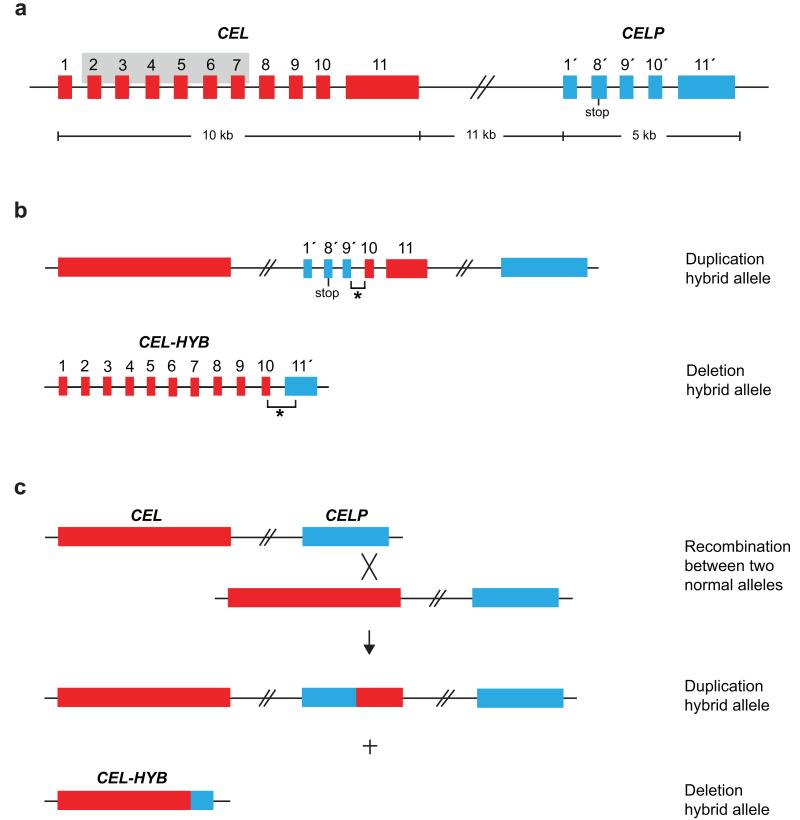
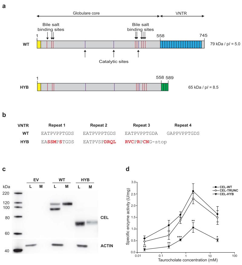
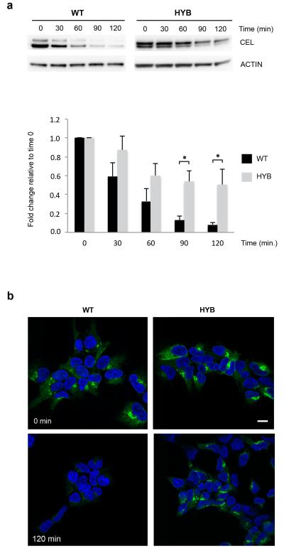
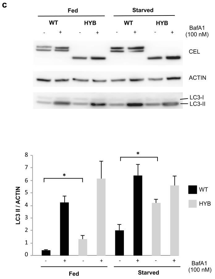
Carboxyl ester lipase is a digestive pancreatic enzyme encoded by the CEL gene. Mutations in CEL cause maturity-onset diabetes of the young as well as pancreatic exocrine dysfunction. Here we describe a hybrid allele (CEL-HYB) originating from a crossover between CEL and its neighboring pseudogene, CELP. In a discovery series of familial chronic pancreatitis cases, we observed CEL-HYB in 14.1% (10/71) of cases compared to 1.0% (5/478) of controls (odds ratio (OR) = 15.5; 95% confidence interval (CI) = 5.1-46.9; P = 1.3 × 10(-6) by two-tailed Fisher's exact test). In three replication studies of nonalcoholic chronic pancreatitis, we identified CEL-HYB in a total of 3.7% (42/1,122) cases and 0.7% (30/4,152) controls (OR = 5.2; 95% CI = 3.2-8.5; P = 1.2 × 10(-11); formal meta-analysis). The allele was also enriched in alcoholic chronic pancreatitis. Expression of CEL-HYB in cellular models showed reduced lipolytic activity, impaired secretion, prominent intracellular accumulation and induced autophagy. These findings implicate a new pathway distinct from the protease-antiprotease system of pancreatic acinar cells in chronic pancreatitis.


Figures

Figure 1
Figure 1
Copy number variants of the human CEL gene. The large red and blue boxes without numbers correspond to the complete CEL and CELP genes, respectively. Smaller boxes represent the individual exons and are numbered 1-11 (CEL) and 1′, 8′-11′ (CELP). The stop codon in exon 8′ of CELP is indicated. (a) Structure of the CEL-CELP gene locus. The grey-shaded area in CEL marks the exon 2-exon 7 region missing in CELP. The size of the locus is shown below the figure; exon and intron sizes are not drawn to scale. (b) Schematic structure of the duplication hybrid allele and the deletion hybrid allele (CEL-HYB) identified in this study. The breakpoint regions (not drawn to scale) are indicated by asterisks. (c) Proposed mechanism of the generation of CEL-HYB by non-allelic homologous recombination between CEL and CELP. The X symbolizes the cross-over event in the exon 10-exon 11 region.
Figure 2
Figure 2
Regional plot showing proxy SNPs of the CEL-HYB variant. R2 represents correlation between the CEL-HYB variant and SNPs based on measured and imputed gene doses. Recombination rate is according to 1,000 Genomes haplotypes Phase I (v3), and reported genes are from Ensembl release 70, January 2013 (see URLs). Genes were limited to biotypes ‘protein-coding’ and ‘pseudogene’. Colors represent levels of R2 with CEL-HYB.
Figure 3
Figure 3
Altered structure, expression and enzyme activity of the CEL-HYB protein compared with wild-type CEL (CEL-WT). (a) Predicted structure of the two protein variants. The yellow box indicates the N-terminal signal peptide. The blue and green boxes correspond to the C-terminal VNTR region of the normal CEL protein and CEL-HYB, respectively. (b) Alignment of the amino acid sequence encoded by the first VNTR repeats of the CEL-WT and CEL-HYB alleles. The altered reading frame of CEL-HYB predicts a change of amino acid sequence starting in the first repeat and a premature stop-codon in repeat 3. Substituted amino acids are shown in red. (c) Expression of CEL-WT and CEL-HYB in stably transfected HEK293 cells. Cell lysates (L) and media (M) were analyzed by Western blotting. Transfection of empty vector (EV) was included as negative control. The actin bands indicate the amount of loaded protein in the lysate lanes only. The data are representative of more than three experiments giving similar result. (d) CEL enzyme activity in conditioned media from stably transfected HEK293 cells. Lipase activity was measured at 37°C in the presence of increasing concentrations of sodium taurocholate as activator. CEL-TRUNC, a variant of the CEL protein completely lacking the C-terminal VNTR, was included for evaluating the effect of the VNTR on enzyme activity. Data are shown as means of seven independent experiments, with statistical comparisons performed between CEL-HYB and CEL-WT activity. Error bars, SEM; *, P = 0.01-0.05; **, P = 0.001-0.01; ***, P < 0.001.
Figure 4
Figure 4
Intracellular properties of CEL-HYB. (a) Reduced cellular clearance of CEL-HYB. Stably transfected HEK293 cells were treated with the protein synthesis inhibitor cycloheximide (1 μg/ml) and examined at the indicated time points. Lysates from cells expressing CEL-WT or CEL-HYB were analyzed for CEL and actin by Western blotting (upper panel). The bands were quantified and CEL level normalized to actin (lower panel). CEL expression at time 0 was arbitrarily set to 1.0. After 120 min exposure to cycloheximide, 50% of CEL-HYB protein was detected in the cell lysate compared with 9% for CEL-WT. Data are shown as means of three independent experiments. Error bars, SEM; *, P = 0.01-0.05. (b) Effect of cycloheximide confirmed by immunostaining and confocal imaging. Cells expressing CEL-HYB showed a much stronger fluorescence signal than CEL-WT-expressing cells after 120 min of cycloheximide treatment. Scale bar, 10 μm. (c) Autophagy induced by CEL-HYB expression. Stably transfected HEK293 cells were incubated in rich (fed) or starvation medium with or without bafilomycin A1 (BafA1) for 3 hours. Cell lysates were analyzed by Western blotting for CEL, actin and the autophagy marker protein LC3 (upper panel). The bands were quantified and LC3–II normalized to actin (lower panel). CEL-HYB-expressing cells grown in full or starved medium showed increased LC3-II levels as compared with CEL-WT-expressing cells. Data are shown as means of three independent experiments. Error bars, SEM; *, P = 0.01-0.05.
Figure 4
Figure 4
Intracellular properties of CEL-HYB. (a) Reduced cellular clearance of CEL-HYB. Stably transfected HEK293 cells were treated with the protein synthesis inhibitor cycloheximide (1 μg/ml) and examined at the indicated time points. Lysates from cells expressing CEL-WT or CEL-HYB were analyzed for CEL and actin by Western blotting (upper panel). The bands were quantified and CEL level normalized to actin (lower panel). CEL expression at time 0 was arbitrarily set to 1.0. After 120 min exposure to cycloheximide, 50% of CEL-HYB protein was detected in the cell lysate compared with 9% for CEL-WT. Data are shown as means of three independent experiments. Error bars, SEM; *, P = 0.01-0.05. (b) Effect of cycloheximide confirmed by immunostaining and confocal imaging. Cells expressing CEL-HYB showed a much stronger fluorescence signal than CEL-WT-expressing cells after 120 min of cycloheximide treatment. Scale bar, 10 μm. (c) Autophagy induced by CEL-HYB expression. Stably transfected HEK293 cells were incubated in rich (fed) or starvation medium with or without bafilomycin A1 (BafA1) for 3 hours. Cell lysates were analyzed by Western blotting for CEL, actin and the autophagy marker protein LC3 (upper panel). The bands were quantified and LC3–II normalized to actin (lower panel). CEL-HYB-expressing cells grown in full or starved medium showed increased LC3-II levels as compared with CEL-WT-expressing cells. Data are shown as means of three independent experiments. Error bars, SEM; *, P = 0.01-0.05.

References

    1. Lombardo D. Bile salt-dependent lipase: its pathophysiological implications. Biochim Biophys Acta. 2001;1533:1–28. - PubMed
    1. Ræder H, et al. Mutations in the CEL VNTR cause a syndrome of diabetes and pancreatic exocrine dysfunction. Nat Genet. 2006;38:54–62. - PubMed
    1. Nilsson J, et al. cDNA cloning of human-milk bile-salt-stimulated lipase and evidence for its identity to pancreatic carboxylic ester hydrolase. Eur J Biochem. 1990;192:543–50. - PubMed
    1. Lidberg U, et al. Genomic organization, sequence analysis, and chromosomal localization of the human carboxyl ester lipase (CEL) gene and a CEL-like (CELL) gene. Genomics. 1992;13:630–40. - PubMed
    1. Madeyski K, Lidberg U, Bjursell G, Nilsson J. Structure and organization of the human carboxyl ester lipase locus. Mamm Genome. 1998;9:334–8. - PubMed

MeSH terms