. 2015 Apr;17(4):524-9.
doi: 10.1038/ncb3134. Epub 2015 Mar 16.

Force transmission during adhesion-independent migration

Affiliations

Force transmission during adhesion-independent migration

Martin Bergert et al. Nat Cell Biol. 2015 Apr.

Abstract

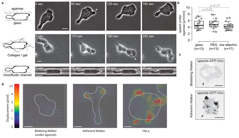
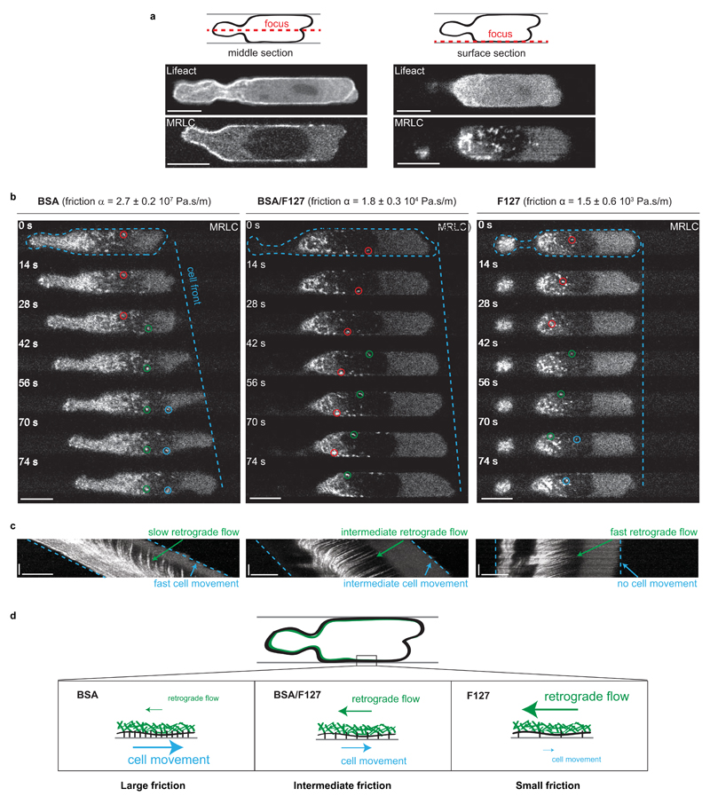
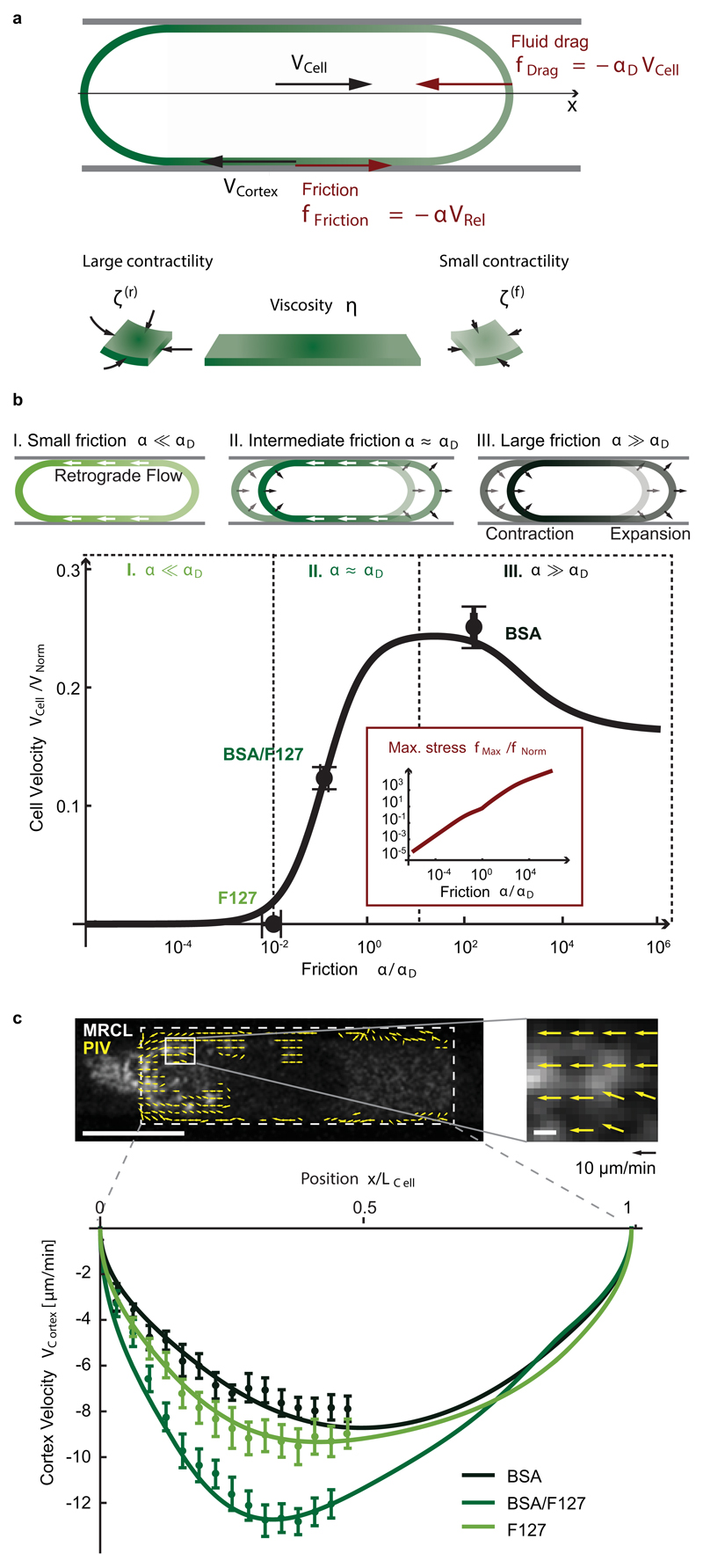
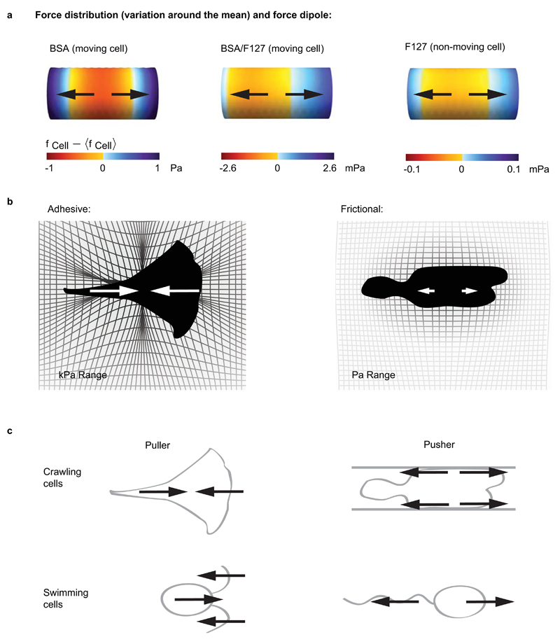
When cells move using integrin-based focal adhesions, they pull in the direction of motion with large, ∼100 Pa, stresses that contract the substrate. Integrin-mediated adhesions, however, are not required for in vivo confined migration. During focal adhesion-free migration, the transmission of propelling forces, and their magnitude and orientation, are not understood. Here, we combine theory and experiments to investigate the forces involved in adhesion-free migration. Using a non-adherent blebbing cell line as a model, we show that actin cortex flows drive cell movement through nonspecific substrate friction. Strikingly, the forces propelling the cell forward are several orders of magnitude lower than during focal-adhesion-based motility. Moreover, the force distribution in adhesion-free migration is inverted: it acts to expand, rather than contract, the substrate in the direction of motion. This fundamentally different mode of force transmission may have implications for cell-cell and cell-substrate interactions during migration in vivo.


Figures

Figure 1
Figure 1. Specific adhesion-independent migration of blebbing Walker cells in confined environments.
a - Timelapses of migrating blebbing Walker cells under agarose, within a three-dimensional collagen-I gel or in a BSA-coated microfluidic channel. Arrowheads: blebs. b - Instantaneous velocities of migrating blebbing Walker cells under agarose on glass, PEG-coated glass and commercial low attachment surfaces (Corning). Cells were manually tracked for 52 min using Fiji. P-Value: Welch's two-sided T-Test; n: number of cells analyzed in 2 independent experiments. Boxes in boxplots extend from the 25th to 75th percentiles, with a line at the median. Whiskers extend to 1.5x IQR (interquartile range) or the max/min datapoints. c - Representative images (inverted contrast) of Walker cells expressing the focal adhesion component speckle GFP-Vinculin. Top: blebbing Walker cell migrating under agarose; no focal adhesions are detected. Bottom: control; clear focal adhesions are formed by adherent Walker cells migrating on glass. Arrows indicate the direction of migration. d - Representative images of in-plane substrate deformations during migration of adherent and non-adherent cells. Non-adherent blebbing Walker cells migrating under agarose on soft (3kPa), elastic PDMS substrates with embedded beads do not elicit substantial bead displacements, while adherent Walker cells and HeLa cells do. Displacement fields caused by the cells were calculated from images of fluorescent beads using a traction force Fiji plugin. Cell outlines are drawn in white. All scale bars: 10 µm.
Figure 2
Figure 2. A minimum friction is required for cortical flow-driven migration of non-adherent Walker cells.
a - F-actin (Lifeact) and Myosin (MRLC) gradients in Walker cells, imaged in the middle cross-section of the cell (left) and within the plane of the actomyosin cortex close to the channel wall (right). Scale bars: 10 µm. b - Timelapses of migrating Walker cells in microchannels with different frictions. The cell substrate friction coefficient α was measured for the 3 different channel coatings (BSA, F127 and BSA/F127-mix) by applying a pressure to the channel entry and measuring the velocity of non-polarized cells (mean ± SEM, see Supplementary Fig. 2 and Materials and Methods for details). Dynamics of the actomyosin cortex in a confocal section at the cell surface were visualized with MRLC-GFP. Colored circles highlight the dynamics of individual myosin foci. Scale bars: 10 µm. c - Kymographs showing the dynamics of the cortex along the long axis of the cell. Scale bars: horizontal: 10 µm, vertical: 50 s. d - Summary of the observed cell behaviors and actomyosin cortex dynamics depending on the friction between the cell and the channel walls.
Figure 3
Figure 3. The mechanics of specific adhesion-independent migration.
a - Schematic and parameters of the physical description of friction-based cell migration. The cell cortex is represented by an axisymmetric surface with viscosity η, subjected to a myosin-II-dependent contractile active tension ζ (see Supplementary Note). A gradient in active tension along the cell axis induces deformations of the cell poles and retrograde cortical flow of velocity VCortex, resulting in cell movement at velocity VCell if the generated friction force fFriction is sufficient to counteract the fluid drag force fDrag. [VRel = VCell + VCortex] b - Top: Cell translocation is achieved by frictional forces resisting a retrograde cortical flow coupled to contraction of the cell rear and expansion of the leading edge. The relative contribution of the two mechanisms depends on the friction coefficient. Bottom: Cell velocity as a function of friction (dots: experimental data (error: SEM), solid line: fitted theoretical curve). Fluid drag αD leads to cell stalling below a threshold value of substrate friction α*/ αD ~ 0.1. The maximum stress exerted on the channel wall (inset) increases for increasing friction, while the cell velocity reaches a plateau. [Normalization: VNorm = (ζ(r) - ζ(f))L/η, fNorm = αD VNorm.] c - Cortical flow profiles in different friction conditions quantified using PIV. Dots: experimental data; lines: fit theoretical curves calculated for measured myosin gradients (Supplementary Fig. 1i and Supplementary Note for details). Scale bars: 10 µm (left panel) and 0.5 µm (right panel). For b/c: nBSA=33, nBSA/F127=25, and nF127=33 cells were analyzed in 5 (BSA), 3 (BSA/F127) and 5 (F127) independent experiments. Data were systematically filtered based on the PIV sample size (error bar: SEM, see Materials and Methods for details).
Figure 4
Figure 4. Force distribution underlying migration in the absence of specific adhesion.
a - Distribution of forces (variation around the mean, see also Supplementary Fig. 4i) exerted by migrating Walker cells on the channel wall. Cell migration direction is to the right, the force is oriented on average in the direction opposite to this motion, and the stress magnitudes are in the mPa-Pa range, considerably smaller than stresses reported for adhesive cellular movement. Propulsive thrust is generated in the rear part of the cells, and cells exert a positive, extensile force dipole on their surrounding environment (ρBSA = 7.7 10-17 N.m, ρBSA/F127 = 2.5 10-18 N.m and ρF127 = 4.4 10-20 N.m). b - Schematic comparison of stresses exerted during adhesive vs. frictional cell migration. Adhesive cells exert large stresses, and induce strong, contractile deformations on their environment; frictional movement relies on small stresses and generates weak, extensile deformations. c - Schematic classification of swimming and crawling cell motion according to the sign of the force dipole.

References

    1. Schwarz US, Safran SA. Physics of adherent cells. Rev Mod Phys. 2013;85:1327–1381.
    1. Lämmermann T, et al. Rapid leukocyte migration by integrin-independent flowing and squeezing. Nature. 2008;453:51–5. - PubMed
    1. Parsons JT, Horwitz AR, Schwartz MA. Cell adhesion: integrating cytoskeletal dynamics and cellular tension. Nat Rev Mol Cell Biol. 2010;11:633–643. - PMC - PubMed
    1. Friedl P. To adhere or not to adhere? Nat Rev Mol Cell Biol. 2010;11:3–3. - PubMed
    1. Renkawitz J, Sixt M. Mechanisms of force generation and force transmission during interstitial leukocyte migration. EMBO Rep. 2010;11:744–50. - PMC - PubMed

Publication types