. 2015 Jun 1;468(2):227-44.
doi: 10.1042/BJ20141021.

RECQ1 interacts with FEN-1 and promotes binding of FEN-1 to telomeric chromatin

Affiliations

RECQ1 interacts with FEN-1 and promotes binding of FEN-1 to telomeric chromatin

Furqan Sami et al. Biochem J. .

Abstract

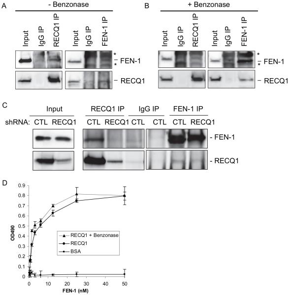
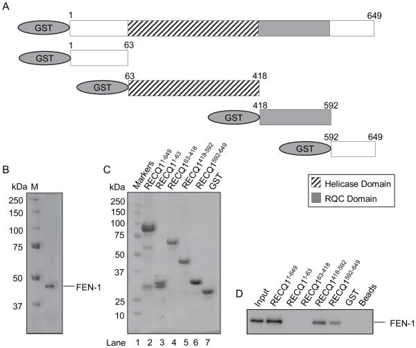
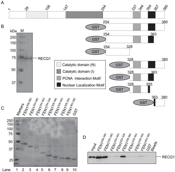
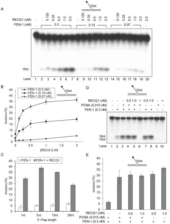
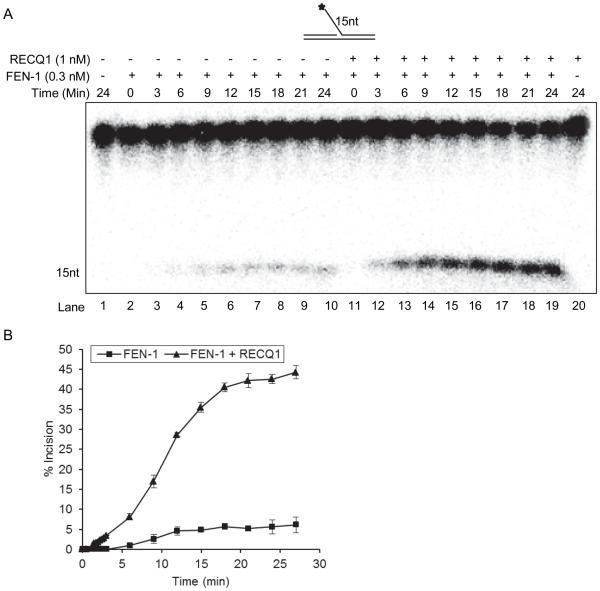
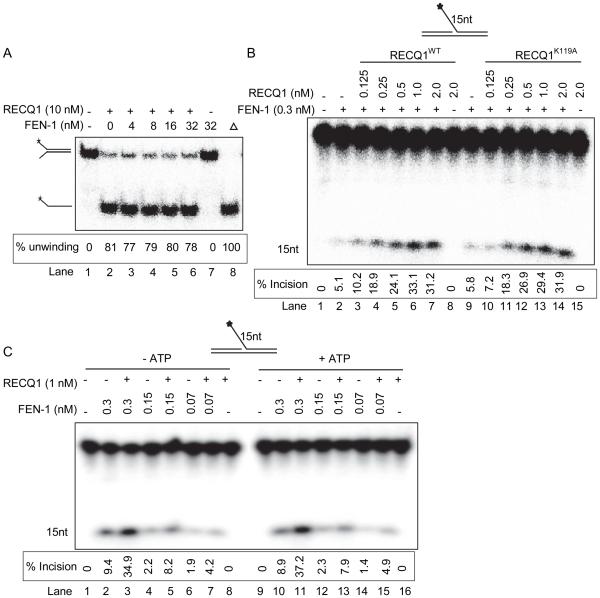
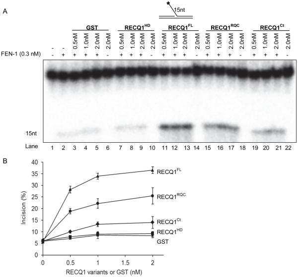
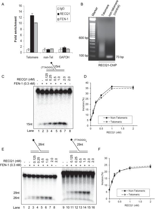
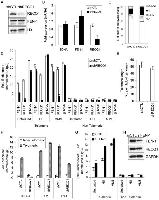
RecQ helicases are a family of highly conserved proteins that maintain genomic stability through their important roles in replication restart mechanisms. Cellular phenotypes of RECQ1 deficiency are indicative of aberrant repair of stalled replication forks, but the molecular functions of RECQ1, the most abundant of the five known human RecQ homologues, have remained poorly understood. We show that RECQ1 associates with FEN-1 (flap endonuclease-1) in nuclear extracts and exhibits direct protein interaction in vitro. Recombinant RECQ1 significantly stimulated FEN-1 endonucleolytic cleavage of 5'-flap DNA substrates containing non-telomeric or telomeric repeat sequence. RECQ1 and FEN-1 were constitutively present at telomeres and their binding to the telomeric chromatin was enhanced following DNA damage. Telomere residence of FEN-1 was dependent on RECQ1 since depletion of RECQ1 reduced FEN-1 binding to telomeres in unperturbed cycling cells. Our results confirm a conserved collaboration of human RecQ helicases with FEN-1 and suggest both overlapping and specialized roles of RECQ1 in the processing of DNA structure intermediates proposed to arise during replication, repair and recombination.


Figures

Figure 1
Figure 1. RECQ1 interacts with FEN-1 in vivo and in vitro
A. Co-IP analysis of RECQ1 interaction with FEN-1 using HeLa nuclear extracts. Immunoprecipitations (IP) with antibodies specific for RECQ1, FEN-1 and preimmune IgG are indicated. Eluted proteins in immunoprecipitate were analyzed by Western blotting and are indicated. RECQ1 IP contained FEN-1. Reciprocal co-IPs of FEN-1 also contained RECQ1. FEN-1 antibody also detected Ig heavy and light chains (indicated by asterisks) in IP-Western and interfered with FEN-1 signal (~ 42 kD). B. Association of RECQ1 and FEN-1 is not mediated via DNA. RECQ1 antibody co-precipitated RECQ1 and FEN-1 using benzonase-treated extract in IP reaction. Reciprocal co-IPs of FEN-1 also contained RECQ1. C. Reciprocal co-IP of RECQ1 and FEN-1 from benzonase-treated extracts prepared from HeLa cells transduced with lentiviral-expressed RNA interference (RNAi) hairpins (shRNA) targeting RECQ1 or luciferase (negative control, CTL). D. Recombinant RECQ1 and FEN-1 proteins directly interact in vitro as shown by ELISA. Either BSA or purified recombinant RECQ1 was coated onto microtiter plates. Following blocking with 3% BSA, appropriate wells were incubated with the indicated concentrations of recombinant FEN-1 (0-50 nM) for 1 h at 30°C. Following washing, RECQ1-bound FEN-1 was detected by ELISA using anti-FEN-1 antibody. The values represent the mean of three independent experiments performed in duplicate with SD indicated by error bars.
Figure 2
Figure 2. FEN-1 binding activity of RECQ1 is contained within RQC and extreme C-terminal end
A. Schematic representation of GST-RECQ1 recombinant fragments used for FEN-1 pull-down experiments. B. Coomassie-stained 10% SDS-PAGE gel showing purified FEN-1 used for binding assays. C. Ponceau S-stained membrane showing protein complexes bound to glutathione sepharose beads in pull-down assay. Beads were mixed with lysate from bacteria expressing GST fusion proteins containing human RECQ1 fragments or GST alone as indicated. D. Purified recombinant FEN-1 protein (200 ng) was added to the indicated GST-RECQ1 bound glutathione sepharose beads. After washing, protein complexes were eluted and resolved by SDS-PAGE. Bound FEN-1 was detected by Western blotting.
Figure 3
Figure 3. RECQ1 binding activity of FEN-1 is contained within amino acids 328-380
A. Schematic representation of GST-FEN-1 recombinant fragments used for pull-down experiments. B. Coomassie-stained 10% SDS-PAGE gel showing purified RECQ1. C. Ponceau S-stained membrane showing protein complexes bound to glutathione sepharose beads in pull-down assay. Beads were mixed with lysate from bacteria expressing GST fusion proteins containing human FEN-1 fragments or GST alone as indicated. D. Purified recombinant RECQ1 protein (200 ng) was added to the indicated GST-FEN-1 bound glutathione sepharose beads. After washing, protein complexes were eluted and resolved by SDS-PAGE. Bound RECQ1 was detected by Western blotting.
Figure 4
Figure 4. RECQ1 stimulates FEN-1 cleavage of 5’-flap DNA substrate
Reactions (20 μl) containing 10 fmol DNA substrate, indicted amounts of FEN-1 and increasing concentration of RECQ1 (0-2.0 nM) were incubated at 37°C for 15 min under conditions described in Experimental methods. Star indicates position of 32P label. A. Phosphorimage of a typical gel of FEN-1 incision activity on a 15 nt 5’-flap DNA substrate. B. Percent incision from the data shown in ‘A’, data points are the mean of three independent experiments with SDs indicated by error bars. C. RECQ1 stimulation of FEN-1 cleavage of 5’-flap substrates with increasing length of 5’-flap (1, 5, 15, and 26 nt). Percent incision by FEN-1 is shown as the mean of three independent experiments with SD indicated by error bars. D. FEN-1 stimulation by PCNA or RECQ1 is mutually exclusive. Incision reactions, performed as above, contained either FEN-1 alone, or the indicated amounts of PCNA or /and RECQ1. E. Percent incision from the representative experiment shown in ‘D’, data points are the mean of three independent experiments with SDs indicated by error bars.
Figure 5
Figure 5. Kinetics of FEN-1 cleavage of the 15nt 5’-flap DNA substrate in the presence or absence of RECQ1
Reactions (200 μl) containing 10 fmol of 15 nt 5’-flap DNA substrate and 0.3 nM of FEN-1 with or without RECQ1 (1 nM) were incubated at 37°C and 20 μl aliquots were removed at indicated time points. A. Phosphorimage of a typical gel from a time course experiment. Increasing times of incubation (0-24 min) for the FEN-1 cleavage reactions conducted in the absence of RECQ1 (lanes 2-10) or in the presence of RECQ1 (lanes 11-19) are indicated. No enzyme and RECQ1 alone reactions are indicated in lane 1 and 20, respectively. B. Percent incision from the data shown in ‘A’, data points are the mean of three independent experiments with SD indicated by error bars.
Figure 6
Figure 6. Stimulation of FEN-1 cleavage of 5’-flap is independent of RECQ1 helicase activity and FEN-1 does not alter RECQ1 helicase activity
A. FEN-1 does not alter RECQ1 helicase activity on a fork duplex. Phosphorimage of a typical helicase gel showing unwinding of a fork duplex by RECQ1 in the presence or absence of FEN-1; heat denatured substrate is indicated by triangle (lane 8). B. Reactions (20 μl) containing 10 fmol of 15nt 5’-flap DNA substrate and indicated concentrations of FEN-1 and/or RECQ1, wild-type (WT) or helicase dead mutant (K119A), proteins were incubated at 37°C for 15 min under standard conditions. Phosphorimage of a typical gel shows helicase-dead mutant of RECQ1 stimulates FEN-1 cleavage of 15nt 5’-flap DNA substrate. Lane 1, no enzyme; lane 2-7, FEN-1+ RECQ1 wild-type (0-2 nM); lane 8-13, FEN-1 + RECQ1 K119A mutant (0-2 nM). C. RECQ1 stimulates FEN-1 activity on 15nt 5’-flap DNA substrate in the absence of ATP. Phosphorimage of a typical gel shows FEN-1 incision products from reactions performed in the presence or absence of ATP.
Figure 7
Figure 7. Mapping of the FEN-1 interaction domains that mediate the functional interaction between RECQ1 and FEN-1
Reactions (20 μl) containing 10 fmol of 15nt 5’-flap DNA substrate, 0.3 nM of FEN-1 and indicated concentrations of GST or GST-fused RECQ1, full-length (FL), helicase domain (HD), RecQ-C-terminus domain (RQC), or C-terminal (Ct), were incubated at 37°C for 15 min under standard conditions. A. Phosphorimage of a typical gel. Lane 1, no enzyme; B. Percent incision from the mean of three independent experiments is shown with SD indicated by error bars.
Figure 8
Figure 8. RECQ1 associates with telomere chromatin and stimulates FEN-1 cleavage of 5’-flap containing telomeric repeat sequence
A. ChIP-qPCR of immunoprecipitated DNA with probes specific for telomeric region. HeLa cells were processed for ChIP using a RECQ1-specific antibody. FEN-1 antibody was used as a positive control for telomere enrichment and rabbit IgG served as negative control in ChIP experiments. Quantification of cross-linked telomere chromatin immunoprecipitated using the indicated antibodies is shown. Fold enrichment over IgG was determined and is shown for each primer pair for the ChIP. Relative occupancy at telomere versus a non-telomere negative control site (DNA containing HBG) and GAPDH shows preferential association of RECQ1 to telomeres. Results are expressed as means ± SEM for at least three independent experiments. B.A representative gel of the amplified telomere DNA immunoprecipitated with RECQ1 antibody. PCR amplified telomere fragments migrated as a smear (50 to ~500 bp). C. Phosphorimage of a typical gel of RECQ1 stimulation of FEN-1 incision of a 15 nt 5’-flap substrate containing TTAGGG repeats in duplex region upstream of 5’-flap. D. Percent incision of 15 nt 5’-flap substrate containing non-telomeric (solid line) or telomeric (dashed line) sequence. Data indicates mean of at least three independent experiments with SD shown as error bars. E. Phosphorimage of a typical gel of RECQ1 (0-2 nM) stimulation of FEN-1 incision of 5’-flap substrates containing non telomeric sequence or TTAGGG repeats in the 5’-flap. F. Percent incision of 5’-flap substrate containing non-telomeric (solid line) or telomeric (dashed line) sequence in the 5’-flap. Data indicates mean of at least three independent experiments with SD shown as error bars.
Figure 9
Figure 9. RECQ1 facilitates constitutive binding of FEN-1 at telomeres
A. Total cell lysates were prepared from stable control-KD (shCTL) and RECQ1-KD (shRECQ1) cells and protein levels of RECQ1 and FEN-1 were measured by Western blotting. Histone H3 serves as loading control. B. RECQ1 silencing and mRNA expression of FEN-1 in control and RECQ1 KD cells was assessed by RT-qPCR normalized to GAPDH. SDHA was included as another housekeeping gene control. C. Cell cycle analysis of control-KD and RECQ1-KD HeLa cells used for ChIP assays. D. Stable control-KD or RECQ1-KD HeLa cells, untreated or treated with HU (2 mM, 16 h) or MMS (15 μg/ml, 16 h), were processed for ChIP using RECQ1, FEN-1, and γH2AX (gH2AX) antibodies. Quantification of telomere chromatin by qPCR of immunoprecipitated DNA with probes specific for telomeric region and a non-telomere negative control site is shown. Fold enrichment over IgG was determined and used to normalize the data to determine specific enrichment of telomere sequence in each case. Results are expressed as means ± SEM for at least three independent experiments. E. Telomere length in control-KD and RECQ1-KD HeLa cells. Results are expressed as means ± SEM for three independent experiments. F. Stable control-KD or RECQ1-KD HeLa cells (untreated) were processed for ChIP-qPCR using indicated antibodies. TRF2 antibody was used as a positive control for telomere enrichment and rabbit IgG served as negative control in ChIP experiments. GAPDH-normalized RT-qPCR quantification of cross-linked telomere chromatin immunoprecipitated using the indicated antibodies is shown. Fold enrichment over IgG was determined and is shown for each primer pair for the ChIP. Relative occupancy at telomere versus a non-telomere negative control site (DNA containing HBG) shows preferential association of RECQ1, FEN-1 and TRF2 to telomeres. As compared to control-KD cells, RECQ1-KD cells show reduced telomere binding of FEN-1 whereas TRF2 binding is comparable. Results are expressed as means ± SEM for at least three independent experiments. G. HeLa cells transfected with control (siCTL) or FEN-1-specific siRNA (siFEN-1), untreated or treated with HU (2 mM, 16 h) or MMS (15 μg/ml, 16 h), were processed for ChIP using RECQ1 and IgG antibodies. Fold enrichment over IgG was determined as described in “D”. Results are expressed as means ± SEM for at least three independent experiments. H. Protein levels of FEN-1 and RECQ1 in total lysates prepared from control or FEN-1 siRNA transfected cells were measured by Western blotting. GAPDH serves as loading control.

References

    1. Sharma S, Doherty KM, Brosh RM., Jr. Mechanisms of RecQ helicases in pathways of DNA metabolism and maintenance of genomic stability. Biochem. J. 2006;398:319–337. - PMC - PubMed
    1. Larsen N, Hickson I. RecQ Helicases: Conserved Guardians of Genomic Integrity. In: Spies M, editor. In DNA Helicases and DNA Motor Proteins. Springer; New York: 2013. pp. 161–184. - PubMed
    1. Brosh RM, Jr., Bohr VA. Human premature aging, DNA repair and RecQ helicases. Nucleic Acids Res. 2007;35:7527–7544. - PMC - PubMed
    1. Brosh RM., Jr. DNA helicases involved in DNA repair and their roles in cancer. Nat. Rev. Cancer. 2013;13:542–558. - PMC - PubMed
    1. Sharma S, Brosh RM., Jr. Unique and important consequences of RECQ1 deficiency in mammalian cells. Cell Cycle. 2008;7:989–1000. - PMC - PubMed

Publication types