. 2015 Mar 16;11(3):e1004736.
doi: 10.1371/journal.ppat.1004736. eCollection 2015 Mar.

A conserved NS3 surface patch orchestrates NS2 protease stimulation, NS5A hyperphosphorylation and HCV genome replication

Affiliations

A conserved NS3 surface patch orchestrates NS2 protease stimulation, NS5A hyperphosphorylation and HCV genome replication

Olaf Isken et al. PLoS Pathog. .

Erratum in

Abstract

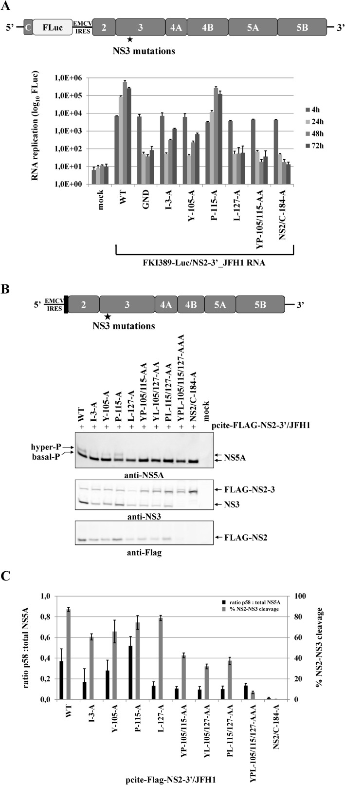
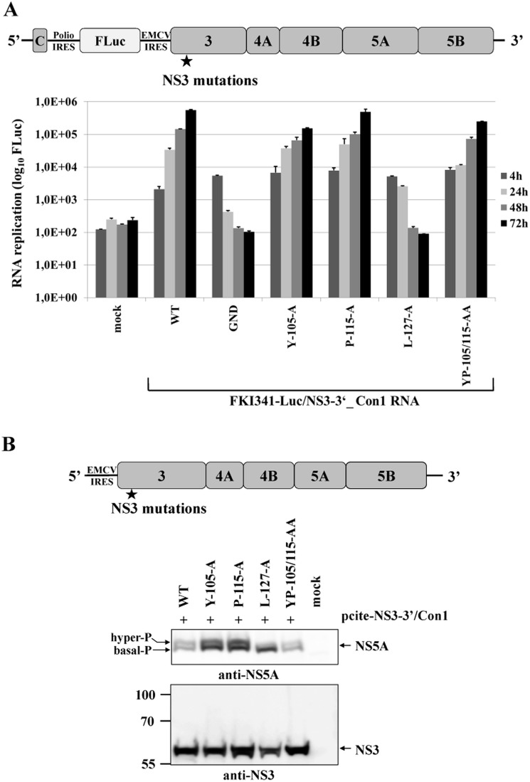
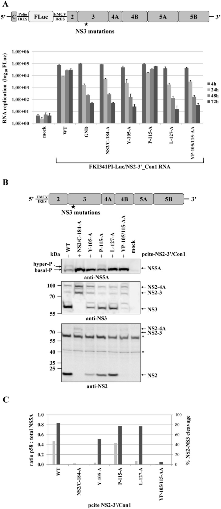
Hepatitis C virus (HCV) infection is a leading cause of liver disease worldwide. The HCV RNA genome is translated into a single polyprotein. Most of the cleavage sites in the non-structural (NS) polyprotein region are processed by the NS3/NS4A serine protease. The vital NS2-NS3 cleavage is catalyzed by the NS2 autoprotease. For efficient processing at the NS2/NS3 site, the NS2 cysteine protease depends on the NS3 serine protease domain. Despite its importance for the viral life cycle, the molecular details of the NS2 autoprotease activation by NS3 are poorly understood. Here, we report the identification of a conserved hydrophobic NS3 surface patch that is essential for NS2 protease activation. One residue within this surface region is also critical for RNA replication and NS5A hyperphosphorylation, two processes known to depend on functional replicase assembly. This dual function of the NS3 surface patch prompted us to reinvestigate the impact of the NS2-NS3 cleavage on NS5A hyperphosphorylation. Interestingly, NS2-NS3 cleavage turned out to be a prerequisite for NS5A hyperphosphorylation, indicating that this cleavage has to occur prior to replicase assembly. Based on our data, we propose a sequential cascade of molecular events: in uncleaved NS2-NS3, the hydrophobic NS3 surface patch promotes NS2 protease stimulation; upon NS2-NS3 cleavage, this surface region becomes available for functional replicase assembly. This model explains why efficient NS2-3 cleavage is pivotal for HCV RNA replication. According to our model, the hydrophobic surface patch on NS3 represents a module critically involved in the temporal coordination of HCV replicase assembly.


Conflict of interest statement

The authors have declared that no competing interests exist.

Figures

Fig 1
Fig 1. Di-alanine scanning mutagenesis of the NS3 protease domain identified amino acids important for the NS2-protease activation by NS3.
(A) The workflow to identify NS3 mutations that inhibit NS2-NS3 cleavage. (B) Scheme of the HCV NS2-NS3 cleavage assay. NS2-mediated cleavage of expressed FLAG-NS2-NS3(1–172)GST/BK polyprotein fragments results in the generation of FLAG-NS2 and NS3(1–172)GST cleavage products. (C) Effect of NS2-NS3 truncations on NS2-NS3 autoprocessing. Plasmids with the indicated NS3 C-terminal truncations were expressed and NS2-NS3 cleavage was detected by Western blotting. NS3(1–180) refers to FLAG-NS2-NS3(1–180)GST/BK. Mock indicates cells transfected with a pcite2a vector control. The positions of FLAG-NS2-NS3-GST, FLAG-NS2 and NS3-GST proteins are indicated by arrows. (D) Levels of NS2-NS3 cleavage were quantified from Western blots. (E) Inhibition of NS2-NS3 cleavage by selected alanine mutations in NS3. Plasmids encoding FLAG-NS2-NS3(1–172)GST with the indicated NS3 mutations were expressed and NS2-NS3 cleavage was detected by Western blotting. WT refers to wild type FLAG-NS2-NS3(1–172), and NS2-C184-A refers to a plasmid expressing NS2-NS3 polyprotein with a NS2-protease active site mutation. Mock indicates cells transfected with a vector control. The positions of the FLAG-NS2-NS3(1–172)GST, NS3(1–172)GST, and FLAG-NS2 proteins are indicated by arrows. (F) Signals of Flag-NS2-NS3GST and NS3GST derivatives were quantified by ImageJ software from three Western blots to calculate the percentage of NS2-NS3 cleavage.
Fig 2
Fig 2. A conserved hydrophobic NS3 surface patch consisting of residues Y105, P115 and L127 is important for NS2-activation by NS3.
(A) Location of the residues I3, Y105, P115 and L127 in the NS3 structure of the genotype 1b. The surface residues are shown in green stick representation. The residues T4, L104, I114 and L126 are depicted in red stick representation. The overall NS3 structure is shown in grey surface representation. Carbon and backbone ribbon are colored in yellow for the protease domain and blue for the helicase domain, respectively. An enlargement of the NS3 surface patch consisting of I3, Y105, P115 and L127 is shown on the right. The figure was generated using Pymol version 1.10 and the coordinates of PDB code 1CU1 [30]. (B) Effect of alanine substitutions in NS3 of HCV strain BK on NS2-NS3 cleavage efficiency. Plasmids expressing either wild type (WT) or mutant FLAG-NS2-NS3(1–172)GST/BK were transfected into Huh7/T7 cells and the cleavage activity was analyzed by Western blot analysis. (C) Quantification of NS2-NS3 cleavage from Western blots related to Fig. 2B using ImageJ software. (D) The hydrophobic character of the NS3 surface area is important for the NS2 protease stimulation by NS3. The cleavage efficiencies of the indicated BK NS2-NS3 polyprotein fragments carrying either charged (arginine) or hydrophobic (phenylalanine) NS3 amino acid substitutions were determined by Western blot analysis. Quantification of these Western blots is presented in S3 Fig. (E) The importance of hydrophobic NS3 surface residues Y105, P115 and L127 for NS2-activation by NS3 is conserved in HCV genotype 2a (JFH1). Plasmids expressing either wild type (WT) or mutant FLAG-NS2-NS3(1–172)GST/JFH1 were transfected into Huh-7/T7 cells and NS2-NS3 cleavage was analyzed by Western blot analysis. Blots shown are representative from three different experiments. (F) Quantification of NS2-NS3 cleavage from Western blots related to Fig. 2E.
Fig 3
Fig 3. Mutational analysis of the NS3-mediated NS2 protease stimulation demonstrates that this process is conserved among different HCV genotypes.
(A) Transient expression of Flag-NS2-3(1–172)GST derivatives from genotype 1b (BK) and genotype 2a (JFH1). After transient expression in the presence of [35S] methionine/cysteine by use of the T7 vaccinia virus system, the cell lysates were subjected to a radioimmunoprecipitation analysis using an anti-GST antibody. The transfected pcite Flag-NS2-NS3(1–172)GST plasmids encoding NS2-3 polyprotein fragments of genotype 1b (BK) or genotype 2a (JFH1) are indicated. WT, wild type; NS2/C184A, NS2-protease inactive mutant; mock, lysates of MVA-T7pol-infected cells were used for RIP assay. The positions of Flag-NS2-NS3(1–172)GST and NS3(1–172)GST are indicated by arrowheads. The positions of the mass standards are shown on the left. (B) Quantification of two independent RIP assays by phosphoimaging.
Fig 4
Fig 4. Mutational analysis of the hydrophobic NS3 surface area in the context of a HCV genotype 2a NS3-5B replicon.
(A) Top, schematic representation of the HCV genotype 2a NS3-5B replicon. Bottom, NS3 mutations were engineered into pFKI389-Luc/NS3-3’_JFH1 (WT), in vitro transcribed RNA was electroporated into Huh7 Lunet cells, and luciferase activity was measured at 4, 24, 48 and 72 h post electroporation (pe). Mean values of three independent experiments are shown. Error bars indicate standard deviations. (B) Top, schematic of the expression construct pcite-NS3-3’/JFH1. This plasmid encodes the NS3 to NS5B sequence of the HCV genotype 2a (JFH1) as indicated. NS3 mutations were introduced into pcite-NS3-3’/JFH1 and plasmids were transfected into Huh-7/T7cells infected with MVA-T7pol vaccinia virus. Bottom, effect of the NS3 mutations on NS3-NS5B polyprotein processing and NS5A hyperphosphorylation. Cell lysates were prepared 20 h post transfection (pt) and analyzed by Western blotting with antibodies directed against HCV NS3, NS4B, NS5A and NS5B. The position of hyper and basally phosphorylated NS5A is indicated on the right. (C) Western blot signals of NS5A p56 and p58 forms of three Western blots were quantified by ImageJ software and the ratio of hyperphosphorylation (p58) to total NS5A was calculated. WT, wild type; GND, polymerase inactive mutant; mock, cells transfected with vector control.
Fig 5
Fig 5. Mutational analysis of the NS3 mutations in the context of a HCV genotype 2a subgenomic NS2-5B replicon revealed NS2-NS3 cleavage is a prerequisite for NS5A hyperphosphorylation.
(A) Top, schematic representation of the FKI389-Luc/NS2-3’_JFH1 replicon. Bottom, the NS3 mutations were introduced into pFKI389-Luc/NS2-3’_JFH1, in vitro transcribed RNA was electroporated into Huh7 Lunet cells, and luciferase activity was measured at 4, 24, 48 and 72 h pe. Mean values of three independent experiments are shown. Error bars indicate standard deviations. (B) Top, schematic of the expression construct pcite-FLAG-NS2-3’/JFH1. This plasmid encodes the T7 promotor sequence fused to the EMCV IRES followed by a FLAG epitope and NS2 to NS5B sequence of the HCV isolate 2a. Indicated NS3-cofactor mutations were engineered into pcite-NS2-3’/JFH1 and plasmids were transfected into Huh-7/T7cells infected with MVA-T7pol vaccinia virus. Bottom, effects of the NS3-cofactor mutations on NS2/3 cleavage, polyprotein processing and NS5A hyperphosphorylation in the context of NS2-5B. Cell lysates were prepared 20 h pt and analyzed by Western blotting with antibodies against FLAG-epitope, NS3 and NS5A. The positions of FLAG-NS2-3, FLAG-NS2, NS3 as well as the hyper and basally phosphorylated NS5A are indicated on the right. (C) Signals of Flag-NS2-3 and NS3 were quantified by ImageJ software from two Western blots to calculate the percentage of NS2-3 cleavage. Signals of NS5A p56 and p58 forms of two Western blots were quantified by ImageJ software and the ratio of hyperphosphorylation (p58) to total NS5A was calculated. WT, wild type; GND, polymerase-inactive mutant; NS2/C-184-A, NS2-protease inactive mutant; mock, cells transfected with vector control.
Fig 6
Fig 6. NS3-L127A inhibits RNA replication and suppresses NS5A hyperphosphorylation in the context of a HCV genotype 1b NS3-5B replicon.
(A) Top, schematic of the HCV genotype 1b NS3-5B replicon. Bottom, the NS3 mutations were engineered into pFKI341-Luc/NS3-3’_Con1 (WT), transcribed RNA was electroporated into Huh7 Lunet cells. Luciferase activity was measured at 4, 24, 48 and 72 h pe. Mean values of three independent experiments are shown, error bars indicate standard deviations. (B) Top, schematic of pcite-NS3-3’/Con1. This plasmid encodes the NS3 to NS5B sequence of the HCV isolate 1b (Con1). NS3 mutations were introduced into pcite-NS3-3’/Con1 and plasmids were transfected into Huh-7/T7cells infected with MVA-T7pol vaccinia virus. Bottom, effects of the NS3 mutations on NS3-5B polyprotein processing and NS5A hyperphosphorylation. Cell lysates were prepared 20 h pt and analyzed by Western blotting with anti NS3 and NS5A antibodies. The position of hyper and basally phosphorylated NS5A is indicated on the left. WT, wild type; GND, polymerase-inactive mutant; mock, cells transfected with vector control.
Fig 7
Fig 7. Mutational analysis of NS3 surface residues in a HCV genotype 1b NS2-5B replicon confirm the critical role of efficient NS2-NS3 cleavage for NS5A hyperphosphorylation.
(A) Top, schematic of the HCV genotype 1b NS2-5B replicon. Bottom, the NS3 mutations were built into pFKI341-Luc/NS2-3’_Con1 and transcribed RNA was electroporated into Huh7 Lunet cells. Luciferase activity was measured at 4, 24, 48 and 72 h pe. Mean values of three independent experiments are shown. Error bars indicate standard deviations. (B) Top, schematic of pcite-NS2-3’/Con1. This plasmid encodes the NS2 to NS5B sequence of the HCV isolate 1b as indicated. NS3 mutations were introduced into pcite-NS2-3’/Con1 and plasmids were transfected into Huh-7/T7cells infected with MVA-T7pol vaccinia virus. Bottom, effects of the NS3 mutations on NS2-5B polyprotein processing and NS5A hyperphosphorylation. Cell lysates were prepared 20 h pt and analyzed by Western blotting with antibodies directed against NS2, NS3 and NS5A. The position of hyper and basally phosphorylated NS5A is indicated. (C) Signals of NS2-3 and NS3 were quantified by ImageJ software from two Western blots to calculate the percentage of NS2-3 cleavage. Signals of NS5A p56 and p58 forms of two Western blots were quantified by ImageJ software and the ratio of hyperphosphorylation (p58) to total NS5A was calculated. WT, wild type; GND, polymerase-inactive mutant; NS2/C-184-A, NS2-protease inactive mutant; mock, lysates from cells transfected with vector control.

Similar articles

Cited by

References

    1. Thomas DL (2013) Global control of hepatitis C: where challenge meets opportunity. Nat Med 19: 850–858. 10.1038/nm.3184 - DOI - PMC - PubMed
    1. McLauchlan J, Lemberg MK, Hope G, Martoglio B (2002) Intramembrane proteolysis promotes trafficking of hepatitis C virus core protein to lipid droplets. EMBO J 21: 3980–3988. - PMC - PubMed
    1. Hijikata M, Kato N, Ootusyama Y, Nakagawa M, Shimotohno K (1991) Gene mapping of the putative structural region of the hepatitis C virus genome by in vitro processing analysis. Proc Natl Acad Sci USA 88: 5547–5551. - PMC - PubMed
    1. Lin C, Lindenbach BD, Prágal BM, McCourt DW, Rice CM (1994) Processing in the Hepatitis C Virus E2-NS2 region: identification of p7 and two distinct E2- specific products with different C termini. J Virol 68: 5063–5073. - PMC - PubMed
    1. Failla C, Tomei L, de Francesco R (1994) Both NS3 and NS4A are required for proteolytic processing of hepatitis C virus nonstructural proteins. J Virol 68: 3753–3760. - PMC - PubMed

Publication types

Substances

LinkOut - more resources