. 2015 Mar 16;10(3):e0119110.
doi: 10.1371/journal.pone.0119110. eCollection 2015.

Identification of protein tyrosine phosphatase receptor gamma extracellular domain (sPTPRG) as a natural soluble protein in plasma

Affiliations

Identification of protein tyrosine phosphatase receptor gamma extracellular domain (sPTPRG) as a natural soluble protein in plasma

Elisabetta Moratti et al. PLoS One. .

Abstract

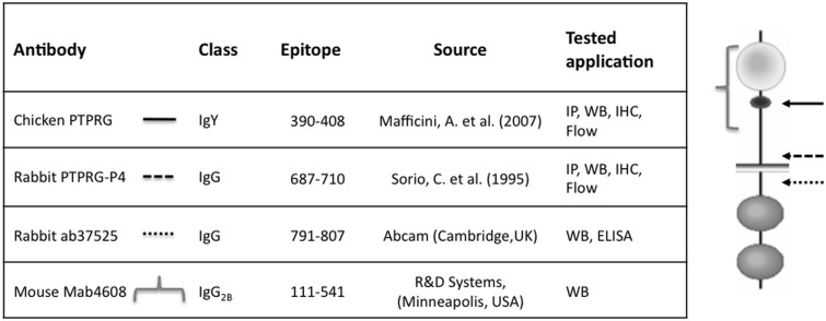
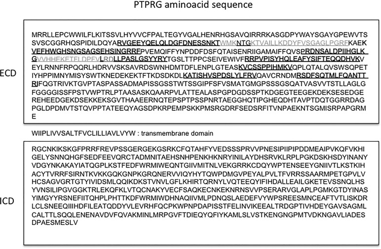
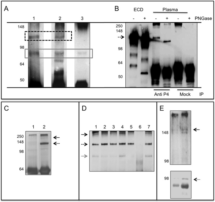
Background: PTPRG is a widely expressed protein tyrosine phosphatase present in various isoforms. Peptides from its extracellular domain have been detected in plasma by proteomic techniques. We aim at characterizing the plasmatic PTPRG (sPTPRG) form and to identify its source.

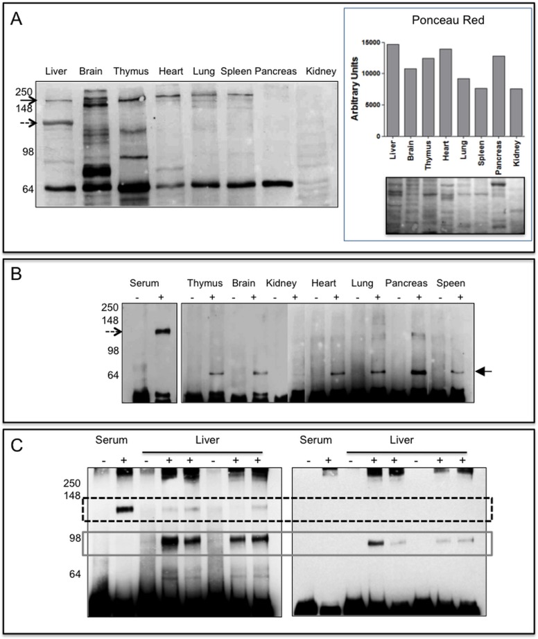
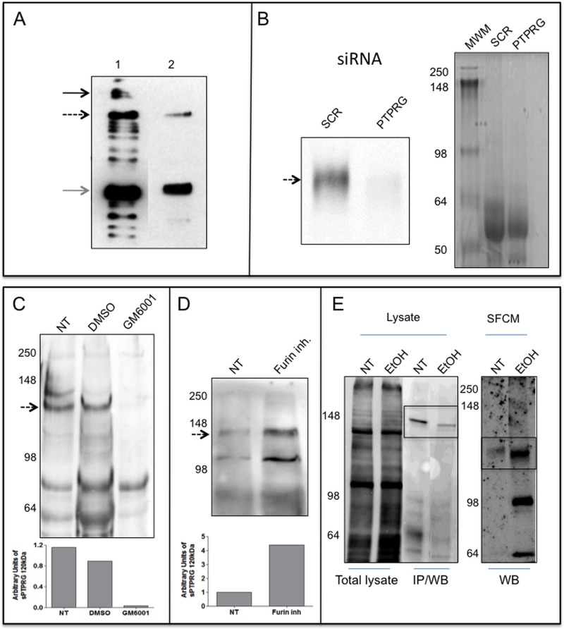
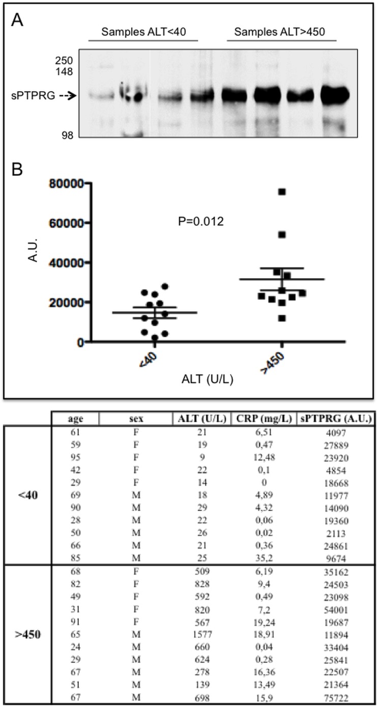
Methodology/principal findings: The expression of sPTPRG was evaluated in human plasma and murine plasma and tissues by immunoprecipitation and Western blotting. The polypeptides identified have an apparent Mr of about 120 kDa (major band) and 90 kDa (minor band) respectively. Full length PTPRG was identified in the 100.000×g pelleted plasma fraction, suggesting that it was present associated to cell-derived vesicles (exosomes). The release of sPTPRG by HepG2 human hepatocellular carcinoma cell line was induced by ethanol and sensitive to metalloproteinase and not to Furin inhibitors. Finally, increased levels of the plasmatic ∼120 kDa isoform were associated with the occurrence of liver damage.

Conclusions: These results demonstrate that sPTPRG represent a novel candidate protein biomarker in plasma whose increased expression is associated to hepatocyte damage. This observation could open a new avenue of investigation in this challenging field.


Conflict of interest statement

Competing Interests: The authors have declared that no competing interests exist.

Figures

Fig 1
Fig 1. Schematic representation of antibodies used.
Epitope: aminoacid number of epitope (NCBI protein accession number P23470), Tested application: IP = immunoprecipitation, WB = Western blot, IHC = immunohystochemistry, Flow = flow cytometry. Right image: approximate position of the epitopes recognized by selected antibody within the predicted protein sequence.
Fig 2
Fig 2. Localization of PTPRG-derived peptides.
Schematic representation of peptides identified by large-scale proteome analysis and reported in the literature[–22]. In grey are the sequences found in all the studies. ECD: extracellular domain, ICD: intracellular domain.
Fig 3
Fig 3. PTPRG plasmatic isoforms.
Panel A: Immunoblotting of human plasma samples immunoprecipitated (IP) with Rb anti-P4 and blotted against: Lane 1: Rb anti-P4, lane 2: mouse monoclonal R&D, lane 3: rabbit polyclonal Abcam. Dashed box: ∼120 kDa PTPRG isoform, gray solid box: ∼90 kDa isoform. Panel B: Immunoblotting with Rb anti-P4 of purified recombinant PTPRG-ECD-Fc lanes 1,2 and immunoprecipitated plasma samples with anti-P4 and RbIgG isotype control (Mock). Sample treated with the deglycosylating enzyme PNGase F are labeled with + sign. Dashed arrow: PTPRG ∼120 kDa isoform. Immunoblotting with Rb anti-P4 of anti-P4 IP human plasma (Panel C) and mouse serum (Panel D) of samples diluted 1:20 in PBS/Tx100 1%. Panel C: Lane 1: Immunoprecipitation with isotype control; Lane, 2: Rb anti-P4. Panel D: Lane 1–5 and 7: IP with Rb anti-P4. Lane 6: IP with RbIgG isotype control. Black arrow: full-length protein, dashed arrow: ∼120 kDa isoform, gray arrow: ∼90 kDa isoform. Left: molecular weight markers. Panel E: Plasma exosomes purified from two individuals were blotted with Rb-anti-P4 (upper panel) and anti ALIX rabbit antibody (lower panel, dotted arrow), an exosome marker. A band corresponding to full-length PTPRG is detectable in both samples (black arrow). Left: molecular weight marker.
Fig 4
Fig 4. PTPRG expression in murine tissues.
Panel A (left): 20μg of lysates from perfused tissues were loaded on 10% SDS-PAGE, separated by gel electrophoresis and then blotted onto a PVDF membrane successively incubated with Rb anti-P4 antibody. Black arrow: full-length protein, dashed arrow highlights the 120 kDa protein detectable in liver lysates. On the right it is reported the densitometric analysis of the same samples stained with Ponceau Red and the corresponding image. Panel B: Immunoblotting reacted with Rb anti-P4 antibody of immunoprecipitated (IP) murine serum and tissue lysates (IP from 250 μg of tissue lysates). Lanes-: IP with Protein G Sepharose rabbit IgG isotype control. Lanes +: IP Protein G Sepharose Rb anti-P4. Dashed arrow: ∼120 kDa isoform, black filled arrow: ∼70 kDa isoform, maybe an intracellular, immature form of a soluble protein, or of a fragment of the full-length protein also present in Panel A. Lack of detectable amounts of full length protein is likely associated to residual proteolysis occurring during the IP step. Panel C: Immunoprecipitation from mouse serum and liver lysates performed with anti P4 antibody (+) or with RbIgG isotype (-). Left: WB performed with Rb anti-P4 reacting against an epitope located within the extracellular domain of human and murine PTPRG. Right: WB with polyclonal anti PTPRG (791–807 Abcam) reacting against an epitope located in the intracellular domain. Dashed box: ∼120 kDa and grey solid box: ∼90 kDa isoforms, respectively.
Fig 5
Fig 5. Expression and release of PTPRG by HepG2 cells.
Panel A: WB with Rb anti-P4 of total lysate and supernatant of HepG2 cell line. Lane 1: 10 μg total cell lysate of HepG2 cell line. Lane 2: serum-free conditioned medium of HepG2 after 100000×g ultra-centrifugation and TCA precipitation. Black arrow: full-length protein, dashed arrow: ∼120 kDa isoform, gray arrow: ∼90 kDa isoform. Panel B: WB with Rb anti-P4 of serum-free conditioned medium of HepG2 cells showing down regulation of sPTPRG by siRNA (siRNA PTPRG) in comparison with a scrambled sequence (SCR). Coomassie blue staining of serum-free conditioned samples demonstrating the loading on comparable amounts of material in both lanes. Panel C: serum-free conditioned medium (SFCM) of HepG2 cells. Left: Molecular weight marker. NT: untreated cells, DMSO: cells treated with vehicle (DMSO), or overnight with 12 μM metalloproteinase inhibitor GM6001 (Ilomastat). Densitometric analysis of sPTPRG 120 kDa isoform related to a common aspecific band approximately at 70 kDa present in all samples (below). Panel D: cells treated overnight with 50 μM furin inhibitor. Dashed arrow: ∼120 kDa isoform. Below is the densitometric analysis expressed as fold increase versus control of the ∼120 kDa band normalized against the total protein load. WB was performed with a chicken anti-PTPRG antibody[27]. Panel E: WB with streptavidin-HRP of cell-surface biotinylated HepG2 cells untreated (NT) or treated (EtOH) for 16 hours with 50 mM ethanol, lysed and immunoprecipitated with anti-P4 antibody (IP/WB, bands boxed). Lower levels of full length PTPRG is present in EtOH treated cells. The same antibody was used in WB analysis on the corresponding serum-free conditioned media precipitated with TCA/acetone (right panel, boxed). The ∼120 kDa PTPRG isoform was detectable in SFCM at higher levels in comparison with untreated cells. The first two lanes represent total cell lysate before IP and demonstrate equal amount of protein and comparable surface biotin labeling of the two samples.
Fig 6
Fig 6. Relationships between liver damage and sPTPRG expression levels in human plasma.
Panel A: representative immunoblotting of plasma (immunoprecipitation with CNBR/anti-P4 and WB with the same antibody) and samples from groups with ALT levels < 40 and > 450 U/L. Dashed arrow indicates the soluble PTPRG isoform of ∼120 kDa (sPTPRG). Panel B: Samples from individuals with Alanine aminotransferase (ALT) levels < 40 and > 450 U/L were evaluated for PTPRG expression measured as normalized values of arbitrary units of light emitted from immunoprecipitated serum samples separated by SDS-PAGE and immunoblotted with anti-P4 antibody (ECL detection system) as described in material and methods. Values of C-reactive protein (CRP, expressed in mg/L) are also shown. Unpaired Student t-test was performed on the data shown. The table summarizes the relevant clinical and biochemical data associated with the samples.

References

    1. Cheung AK, Lung HL, Hung SC, Law EW, Cheng Y, Yau WL, et al. Functional analysis of a cell cycle-associated, tumor-suppressive gene, protein tyrosine phosphatase receptor type G, in nasopharyngeal carcinoma. Cancer Res. 2008; 68: 8137–8145. 10.1158/0008-5472.CAN-08-0904 - DOI - PubMed
    1. Wang Z, Shen D, Parsons DW, Bardelli A, Sager J, Szabo S, et al. Mutational analysis of the tyrosine phosphatome in colorectal cancers. Science. 2004; 304: 1164–1166. - PubMed
    1. Panagopoulos I, Pandis N, Thelin S, Petersson C, Mertens F, Borg A, et al. The FHIT and PTPRG genes are deleted in benign proliferative breast disease associated with familial breast cancer and cytogenetic rearrangements of chromosome band 3p14. Cancer Res. 1996; 56: 4871–4875. - PubMed
    1. Lamprianou S, Vacaresse N, Suzuki Y, Meziane H, Buxbaum JD, Schlessinger J, et al. Receptor protein tyrosine phosphatase gamma is a marker for pyramidal cells and sensory neurons in the nervous system and is not necessary for normal development. Mol Cell Biol. 2006; 26: 5106–5119. - PMC - PubMed
    1. Zhang W, Savelieva KV, Tran DT, Pogorelov VM, Cullinan EB, Baker KB, et al. Characterization of PTPRG in knockdown and phosphatase-inactive mutant mice and substrate trapping analysis of PTPRG in mammalian cells. PLoS One. 2012; 7: e45500 10.1371/journal.pone.0045500 - DOI - PMC - PubMed

Publication types

LinkOut - more resources