

High-resolution structure of the Escherichia coli ribosome
- PMID: 25775265
- PMCID: PMC4429131
- DOI: 10.1038/nsmb.2994
High-resolution structure of the Escherichia coli ribosome
Abstract
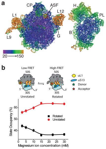
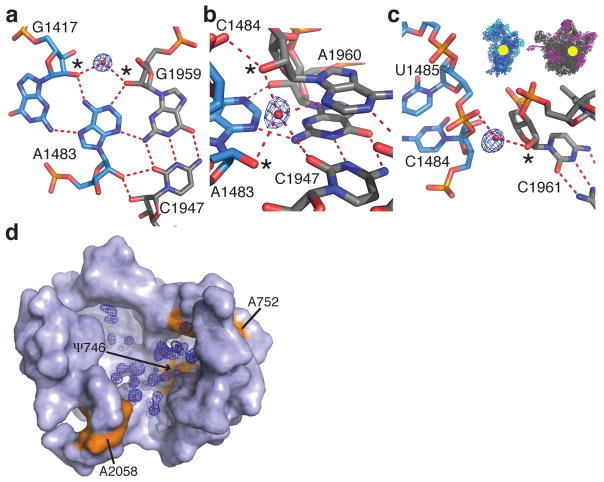
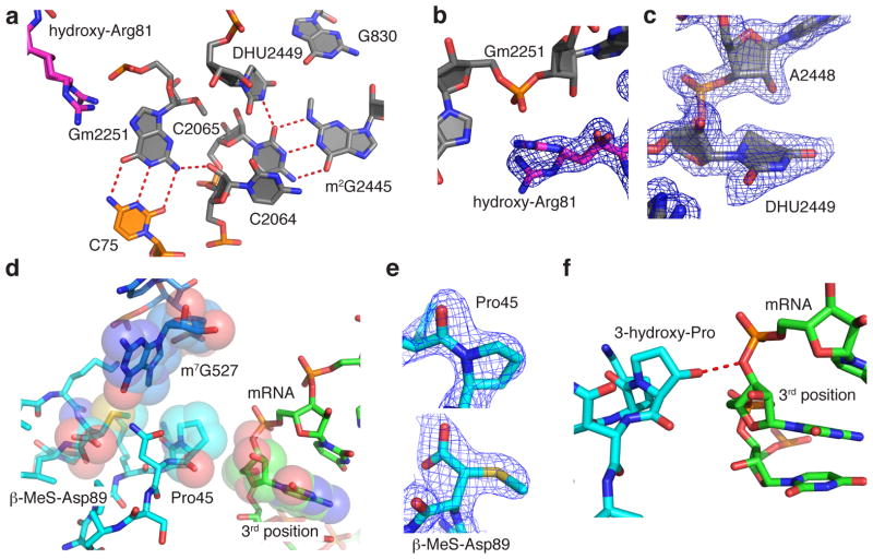
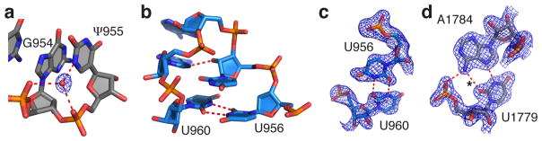
Protein synthesis by the ribosome is highly dependent on the ionic conditions in the cellular environment, but the roles of ribosome solvation have remained poorly understood. Moreover, the functions of modifications to ribosomal RNA and ribosomal proteins have also been unclear. Here we present the structure of the Escherichia coli 70S ribosome at 2.4-Å resolution. The structure reveals details of the ribosomal subunit interface that are conserved in all domains of life, and it suggests how solvation contributes to ribosome integrity and function as well as how the conformation of ribosomal protein uS12 aids in mRNA decoding. This structure helps to explain the phylogenetic conservation of key elements of the ribosome, including post-transcriptional and post-translational modifications, and should serve as a basis for future antibiotic development.
Figures
Similar articles
-
Structure of the bacterial ribosome at 2 Å resolution.Elife. 2020 Sep 14;9:e60482. doi: 10.7554/eLife.60482. Elife. 2020. PMID: 32924932 Free PMC article.
-
X-ray crystal structures of the WT and a hyper-accurate ribosome from Escherichia coli.Proc Natl Acad Sci U S A. 2003 Jul 22;100(15):8682-7. doi: 10.1073/pnas.1133380100. Epub 2003 Jul 9. Proc Natl Acad Sci U S A. 2003. PMID: 12853578 Free PMC article.
-
Structural basis for the interaction of protein S1 with the Escherichia coli ribosome.Nucleic Acids Res. 2015 Jan;43(1):661-73. doi: 10.1093/nar/gku1314. Epub 2014 Dec 15. Nucleic Acids Res. 2015. PMID: 25510494 Free PMC article.
-
Deepening ribosomal insights.ACS Chem Biol. 2006 Oct 24;1(9):567-9. doi: 10.1021/cb600407u. ACS Chem Biol. 2006. PMID: 17168551 Review.
-
Insights into protein biosynthesis from structures of bacterial ribosomes.Curr Opin Struct Biol. 2007 Jun;17(3):302-9. doi: 10.1016/j.sbi.2007.05.009. Epub 2007 Jun 15. Curr Opin Struct Biol. 2007. PMID: 17574829 Review.
Cited by
-
Dynamic contact network between ribosomal subunits enables rapid large-scale rotation during spontaneous translocation.Nucleic Acids Res. 2015 Aug 18;43(14):6747-60. doi: 10.1093/nar/gkv649. Epub 2015 Jun 24. Nucleic Acids Res. 2015. PMID: 26109353 Free PMC article.
-
Structure of the bacterial ribosome at 2 Å resolution.Elife. 2020 Sep 14;9:e60482. doi: 10.7554/eLife.60482. Elife. 2020. PMID: 32924932 Free PMC article.
-
Computationally-guided design and selection of high performing ribosomal active site mutants.Nucleic Acids Res. 2022 Dec 9;50(22):13143-13154. doi: 10.1093/nar/gkac1036. Nucleic Acids Res. 2022. PMID: 36484094 Free PMC article.
-
Genome-wide RNAi screen identifies novel players in human 60S subunit biogenesis including key enzymes of polyamine metabolism.Nucleic Acids Res. 2022 Mar 21;50(5):2872-2888. doi: 10.1093/nar/gkac072. Nucleic Acids Res. 2022. PMID: 35150276 Free PMC article.
-
Force measurements show that uL4 and uL24 mechanically stabilize a fragment of 23S rRNA essential for ribosome assembly.RNA. 2019 Apr;25(4):472-480. doi: 10.1261/rna.067504.118. Epub 2019 Jan 31. RNA. 2019. PMID: 30705137 Free PMC article.
References
Publication types
MeSH terms
Substances
Associated data
- Actions
Grants and funding
LinkOut - more resources
Full Text Sources
Other Literature Sources
