

Age, Sex, and APOE ε4 Effects on Memory, Brain Structure, and β-Amyloid Across the Adult Life Span
- PMID: 25775353
- PMCID: PMC4428984
- DOI: 10.1001/jamaneurol.2014.4821
Age, Sex, and APOE ε4 Effects on Memory, Brain Structure, and β-Amyloid Across the Adult Life Span
Abstract
Importance: Typical cognitive aging may be defined as age-associated changes in cognitive performance in individuals who remain free of dementia. Ideally, the full adult age spectrum should be included to assess brain imaging findings associated with typical aging.
Objective: To compare age, sex, and APOE ε4 effects on memory, brain structure (adjusted hippocampal volume [HVa]), and amyloid positron emission tomography (PET) in cognitively normal individuals aged 30 to 95 years old.
Design, setting, and participants: Cross-sectional observational study (March 2006 to October 2014) at an academic medical center. We studied 1246 cognitively normal individuals, including 1209 participants aged 50 to 95 years old enrolled in a population-based study of cognitive aging and 37 self-selected volunteers aged 30 to 49 years old.
Main outcomes and measures: Memory, HVa, and amyloid PET.
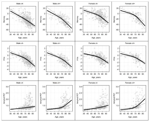
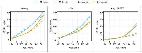
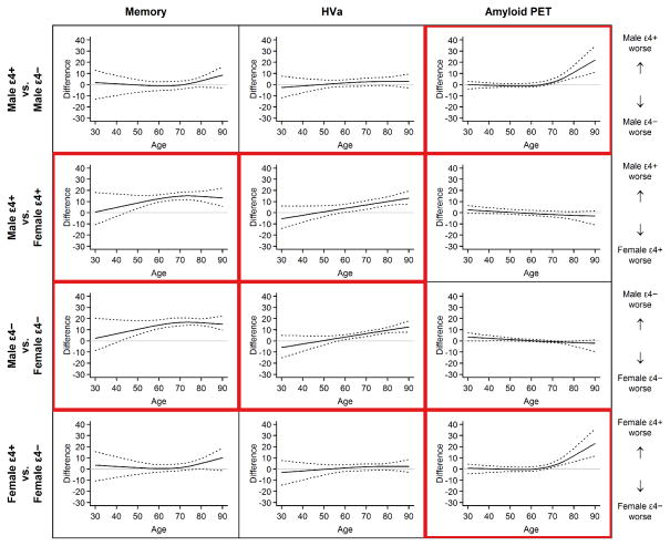
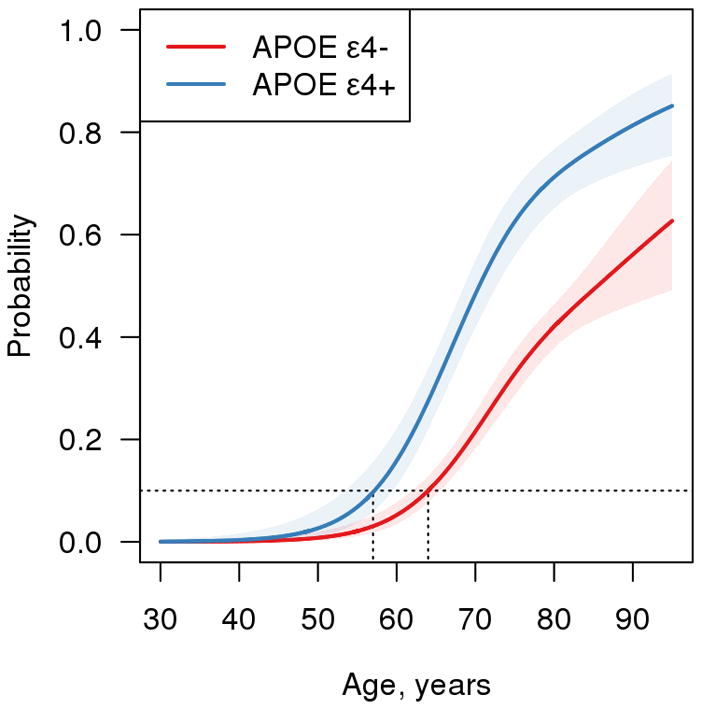
Results: Overall, memory worsened from age 30 years through the 90s. The HVa worsened gradually from age 30 years to the mid-60s and more steeply beyond that age. The median amyloid PET was low until age 70 years and increased thereafter. Memory was worse in men than in women overall (P < .001) and more specifically beyond age 40 years. The HVa was lower in men than in women overall (P < .001) and more specifically beyond age 60 years. There was no sex difference in amyloid PET at any age. Within each sex, memory performance and HVa were not different by APOE ε4 status at any age. From age 70 years onward, APOE ε4 carriers had significantly greater median amyloid PET than noncarriers. However, the ages at which 10% of the population were amyloid PET positive were 57 years for APOE ε4 carriers and 64 years for noncarriers.
Conclusions and relevance: Male sex is associated with worse memory and HVa among cognitively normal individuals, while APOE ε4 is not. In contrast, APOE ε4 is associated with greater amyloid PET (from age 70 years onward), while sex is not. Worsening memory and HVa occur at earlier ages than abnormal amyloid PET. Therefore, neuropathological processes other than β-amyloidosis must underlie declines in brain structure and memory function in middle age. Our findings are consistent with a model of late-onset Alzheimer disease in which β-amyloidosis arises in later life on a background of preexisting structural and cognitive decline that is associated with aging and not with β-amyloid deposits.
Figures
Comment in
-
A call for new thoughts about what might influence human brain aging: aging, apolipoprotein E, and amyloid.JAMA Neurol. 2015 May;72(5):500-2. doi: 10.1001/jamaneurol.2015.33. JAMA Neurol. 2015. PMID: 25775040 Free PMC article. No abstract available.
References
-
- Villemagne VL, Burnham S, Bourgeat P, et al. Amyloid beta deposition, neurodegeneration, and cognitive decline in sporadic Alzheimer’s disease: a prospective cohort study. Lancet Neurol. 2013;12(4):357–367. - PubMed
Publication types
MeSH terms
Substances
Grants and funding
- R01-AG041851/AG/NIA NIH HHS/United States
- R01 AG041851/AG/NIA NIH HHS/United States
- R01 AG034676/AG/NIA NIH HHS/United States
- P50 AG016574/AG/NIA NIH HHS/United States
- U01 AG024904/AG/NIA NIH HHS/United States
- K12 HD065987/HD/NICHD NIH HHS/United States
- R01 AG040042/AG/NIA NIH HHS/United States
- R01 AG011378/AG/NIA NIH HHS/United States
- R01 AG037551/AG/NIA NIH HHS/United States
- R01 AG043392/AG/NIA NIH HHS/United States
- R01 AG032990/AG/NIA NIH HHS/United States
- P50 AG044170/AG/NIA NIH HHS/United States
- R01-AG011378/AG/NIA NIH HHS/United States
- U01-AG06786/AG/NIA NIH HHS/United States
- U01 AG006786/AG/NIA NIH HHS/United States
LinkOut - more resources
Full Text Sources
Other Literature Sources
Medical
Miscellaneous
