. 2015 Mar 16;11(3):e1004682.
doi: 10.1371/journal.ppat.1004682. eCollection 2015 Mar.

A crystal structure of the Dengue virus NS5 protein reveals a novel inter-domain interface essential for protein flexibility and virus replication

Affiliations

A crystal structure of the Dengue virus NS5 protein reveals a novel inter-domain interface essential for protein flexibility and virus replication

Yongqian Zhao et al. PLoS Pathog. .

Abstract

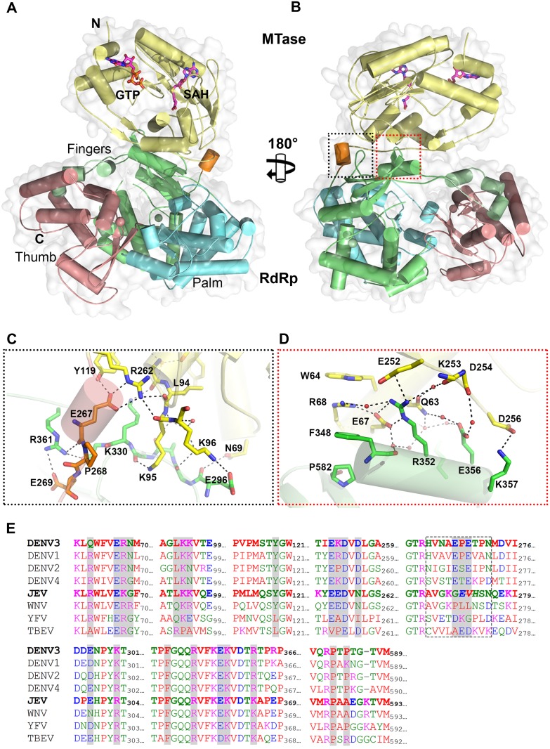
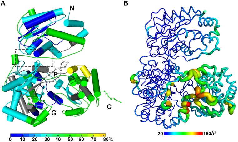
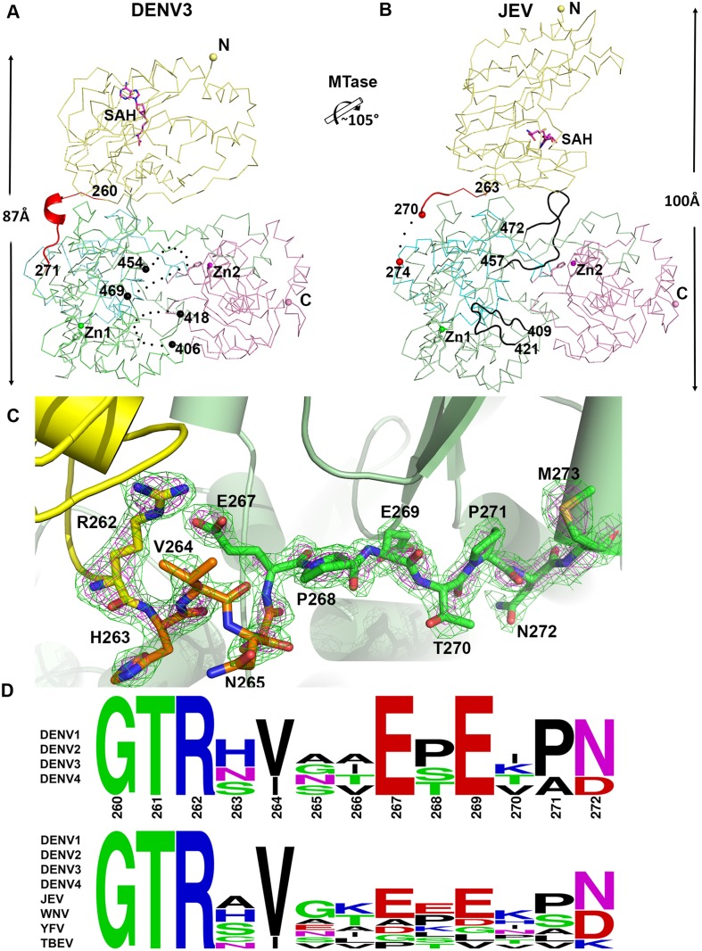
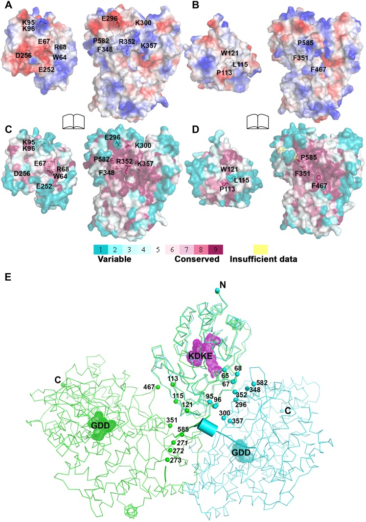
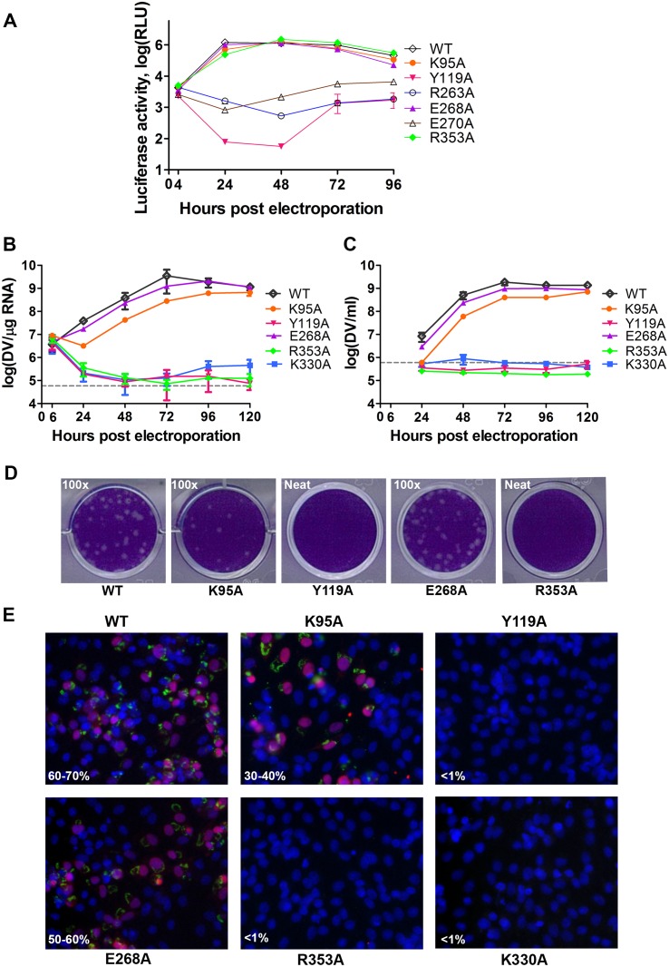
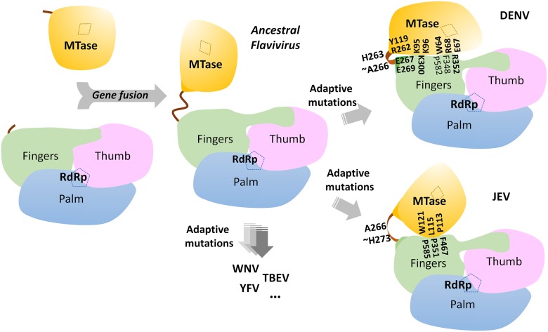
Flavivirus RNA replication occurs within a replication complex (RC) that assembles on ER membranes and comprises both non-structural (NS) viral proteins and host cofactors. As the largest protein component within the flavivirus RC, NS5 plays key enzymatic roles through its N-terminal methyltransferase (MTase) and C-terminal RNA-dependent-RNA polymerase (RdRp) domains, and constitutes a major target for antivirals. We determined a crystal structure of the full-length NS5 protein from Dengue virus serotype 3 (DENV3) at a resolution of 2.3 Å in the presence of bound SAH and GTP. Although the overall molecular shape of NS5 from DENV3 resembles that of NS5 from Japanese Encephalitis Virus (JEV), the relative orientation between the MTase and RdRp domains differs between the two structures, providing direct evidence for the existence of a set of discrete stable molecular conformations that may be required for its function. While the inter-domain region is mostly disordered in NS5 from JEV, the NS5 structure from DENV3 reveals a well-ordered linker region comprising a short 310 helix that may act as a swivel. Solution Hydrogen/Deuterium Exchange Mass Spectrometry (HDX-MS) analysis reveals an increased mobility of the thumb subdomain of RdRp in the context of the full length NS5 protein which correlates well with the analysis of the crystallographic temperature factors. Site-directed mutagenesis targeting the mostly polar interface between the MTase and RdRp domains identified several evolutionarily conserved residues that are important for viral replication, suggesting that inter-domain cross-talk in NS5 regulates virus replication. Collectively, a picture for the molecular origin of NS5 flexibility is emerging with profound implications for flavivirus replication and for the development of therapeutics targeting NS5.


Conflict of interest statement

We have read the journal’s policy and have the following conflicts: TSS, SPL and PYS are currently or have been under the employment of Novartis, which provided funds and equipment to conduct the studies and where they receive salary, benefits and stock. This does not alter our adherence to all PLOS policies on sharing data and materials.

Figures

Fig 1
Fig 1. Crystal structure of DENV3 NS5.
(A) Overall structure of the NS5 protein from DENV3 in cartoon representation viewing from the bottom of RdRp. MTase is in yellow, RdRp fingers in green, palm in blue, thumb in salmon. The linker helix (residues 263–267) between the two domains is in orange. GTP and co-factor SAH are shown as sticks and labelled. Zinc ions are shown as spheres. (B) View of the NS5 molecule from the top of RdRp, which is rotated by 180° around a vertical axis as in (A). Interface regions are boxed. (C) and (D) Close-up views of the interface between the MTase domain and RdRp domain as indicated in (B). Key residues for inter-domain interactions are shown as sticks and labeled. (E) Multiple sequence alignment of flavivirus NS5 proteins. Interface residues are highlighted in gray. The linker residues (263–272 in DENV3 NS5) are boxed. List of accession numbers for genes and proteins used for alignment: DENV3: gi|50347097|gb|AAT75224.1|; DENV1: gi|194338413|gb|ACF49259.1|; DENV2: gi|266813|sp|P29990.1|; DENV4: gi|425895219|gb|AFY10034.1|; JEV: gi|4416167|gb|AAD20233.1|; WNV: gi|607369775|gb|AHW48802.1|; YFV: gi|27735297|ref|NP_776009.1|; TBEV: gi|1709707|sp|Q01299.1|.
Fig 2
Fig 2. The dynamics of NS5 in solution probed by HDX-MS.
(A) Heat map data overlaid onto a crystal structure for the DENV3 NS5. The color key indicates the extent of the deuterium uptake (%), in which blue means the lowest deuterium uptake. Peptides in gray are regions where information of deuterium uptake is missing. F: motif F; G: motif G. (B) Putty cartoon view of temperature factor variation on the crystal structure of DENV3 NS5, colored from low to high values (20~180 Å2 as blue to red).
Fig 3
Fig 3. Comparison of conformations between DENV3 NS5 and JEV NS5 structures.
(A) and (B) Side-by-side view of the DENV3 NS5 and the JEV NS5 structures in thin ribbon style. Same color scheme as in Fig. 1: MTase is in yellow, RdRp fingers in green, palm in blue, thumb in salmon. The linker regions (260–271 in DENV3 NS5; 263–274 in JEV NS5) are shown as cartoon in red. Missing residues (407–417 and 455–468 in DENV3 NS5; 271–273 in JEV NS5) are indicated as dots. The rotation axis that relates both MTase domains is indicated with the corresponding angle. (C) The simulated annealing mFobs-DFcalc omit maps are in green, contoured at 3σ and in magenta, contoured at 5σ. (D) Sequence conservation of the linker region of DENV 1–4 and representative flaviviruses. Figure is generated with WebLogo [71].
Fig 4
Fig 4. Comparison of interfaces between DENV3 and JEV NS5.
RdRp domains of the two structures have been displayed in the same orientation and the buried surfaces (indicated by the labeled interface residues) from the RdRp domains are relatively overlapping between the two NS5 structures. (A) and (B) Analysis of the electrostatic properties of the domain interfaces of the two NS5 structures. Positive surface charge is highlighted in red, negative charge in blue. (C) and (D) Analysis of the evolutionary conservation of the domain interfaces of the two NS5 structures. The color key indicates the degree of conservation, with cyan means highly variable and purple means highly conserved. (E) The relative orientations of the RdRp domain relative to the MTase domain in two structures in thin ribbon style. DENV3 NS5 is in blue, JEV NS5 is in green. “KDKE” highlights the MTase active site shown as mesh in purple and “GDD” for the RdRp active site shown as mesh. Residues at interface are highlighted and shown as dots. Linker (262–272) in DENV3 NS5 is shown as cartoon.
Fig 5
Fig 5. Replication profiles of NS5 interface mutants.
(A) Renilla luciferase activities of DENV4 WT and mutant replicons. Equal amounts of replicon RNA (WT or mutants) were electroporated into BHK-21 cells. At the indicated time points, the transfected cells were lysed and assayed for luciferase activities. The y axis shows the log10 value of Renilla luciferase activity (RLU). Each data point is the average for three replicates, and error bars show the standard deviations. (B) 10μg in vitro transcribed infectious clone RNA was electroporated into BHK-21 cells and viral replication was monitored over a course of 5 days. Intracellular viral RNA replication as detected by qRT-PCR. The grey dotted line represents the background detection of uninfected cells. (C) Extracellular viral RNA in the supernatants detected by qRT-PCR. The grey dotted line denotes background signal of uninfected supernatant. (D) Plaque morphologies of WT and the mutants at 72 hours post electroporation. (E) IFA images showing dsRNA and NS5 co-staining and percentage infection of cells at 72 hours post electroporation.
Fig 6
Fig 6. A schematic model for the divergent evolution of flaviviral NS5 proteins.
The same color scheme as in Fig. 1 is used: MTase is in yellow, RdRp fingers in green, palm in blue, thumb in salmon. The linker region 310 helix (residues 263–266) between the two domains is in orange. Active sites for MTase and RdRp are labelled with dotted tetragon and pentagon respectively. Linker residues and interface residues are labeled. A possible evolutionary pathway is presented: the MTase domain and RdRp domain originally existing as two separate proteins (left) became linked together to form the NS5 protein from an ancestral Flavivirus, possibly through gene fusion. This fusion promoted colocalization of both enzymatic activities and effectively increased the effective concentration of the proteins with respect to each other (middle panel). Following further (divergent) evolution, NS5 acquired different adaptive mutations and gave rise to the NS5 proteins now observed for various viruses, including DENV, JEV and possibly other flaviviruses) (right panel). Thus NS5 proteins from DENV and JEV may have different conformations and different allosteric mechanisms, in which the MTase and RdRp domain cross-talk to each other through unique interfaces specific to either DENV or JEV [72].

References

    1. Bhatt S, Gething PW, Brady OJ, Messina JP, Farlow AW, et al. (2013) The global distribution and burden of dengue. Nature 496: 504–507. 10.1038/nature12060 - DOI - PMC - PubMed
    1. Mackenzie J (2005) Wrapping things up about virus RNA replication. Traffic 6: 967–977. - PMC - PubMed
    1. Miller S, Krijnse-Locker J (2008) Modification of intracellular membrane structures for virus replication. Nat Rev Microbiol 6: 363–374. 10.1038/nrmicro1890 - DOI - PMC - PubMed
    1. Salonen A, Ahola T, Kaariainen L (2005) Viral RNA replication in association with cellular membranes. Curr Top Microbiol Immunol 285: 139–173. - PMC - PubMed
    1. Egloff MP, Benarroch D, Selisko B, Romette JL, Canard B (2002) An RNA cap (nucleoside-2′-O-)-methyltransferase in the flavivirus RNA polymerase NS5: crystal structure and functional characterization. EMBO J 21: 2757–2768. - PMC - PubMed

Publication types

MeSH terms

Substances

LinkOut - more resources