. 2015 May;35(10):1871-81.
doi: 10.1128/MCB.01521-14. Epub 2015 Mar 16.

The Human RNA Polymerase I Transcription Terminator Complex Acts as a Replication Fork Barrier That Coordinates the Progress of Replication with rRNA Transcription Activity

Affiliations

The Human RNA Polymerase I Transcription Terminator Complex Acts as a Replication Fork Barrier That Coordinates the Progress of Replication with rRNA Transcription Activity

Yufuko Akamatsu et al. Mol Cell Biol. 2015 May.

Abstract

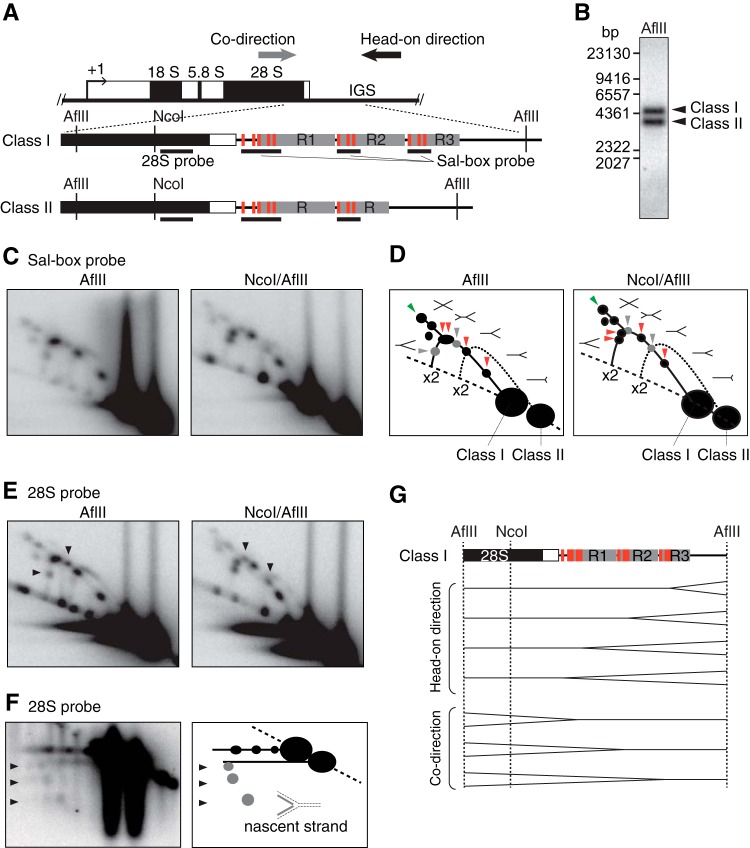
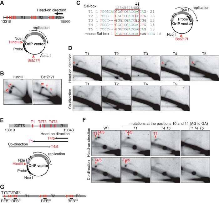
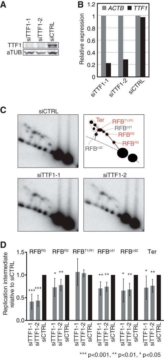
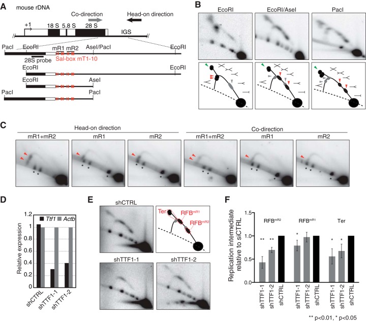
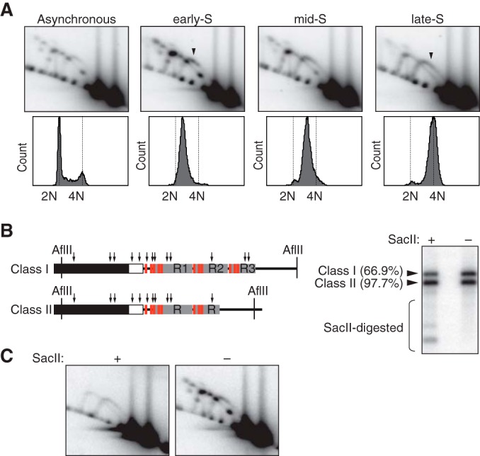
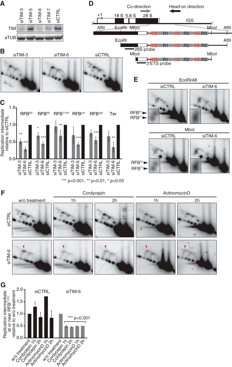
In S phase, the replication and transcription of genomic DNA need to accommodate each other, otherwise their machineries collide, with chromosomal instability as a possible consequence. Here, we characterized the human replication fork barrier (RFB) that is present downstream from the 47S pre-rRNA gene (ribosomal DNA [rDNA]). We found that the most proximal transcription terminator, Sal box T1, acts as a polar RFB, while the other, Sal box T4/T5, arrests replication forks bidirectionally. The fork-arresting activity at these sites depends on polymerase I (Pol I) transcription termination factor 1 (TTF-1) and a replisome component, TIMELESS (TIM). We also found that the RFB activity was linked to rDNA copies with hypomethylated CpG and coincided with the time that actively transcribed rRNA genes are replicated. Failed fork arrest at RFB sites led to a slowdown of fork progression moving in the opposite direction to rRNA transcription. Chemical inhibition of transcription counteracted this deceleration of forks, indicating that rRNA transcription impedes replication in the absence of RFB activity. Thus, our results reveal a role of RFB for coordinating the progression of replication and transcription activity in highly transcribed rRNA genes.


Figures

FIG 1
FIG 1
Human cells contain multiple RFB sites near Sal boxes downstream from the 47S pre-rRNA-coding region. (A) Structure of human rDNA. AflII fragments containing the transcription terminator region with its upstream coding region are magnified, showing Sal boxes (red bars), R repeats (gray rectangles), the positions of 28S or Sal box probes (black lines), and the directions of replication (arrows). Class I and class II rDNAsw differ in the number of R repeats. (B) Digestion of genomic DNA from HeLa cells with AflII reveals two restriction length variations, class I and class II, that, after Southern blotting, were visualized by hybridization with the 28S probe. (C) 2-D gel analysis of replication intermediates on AflII and AflII/NcoI fragments visualized with the Sal box probe. Note that multiple Y forks accumulate on the Y arc of class I but not on that of class II. (D) Schematic diagram of results shown in panel C. Shapes of replication intermediates and Y-, double-Y-, and X-shaped molecules are represented. Nonreplicating molecules appear on the dashed line. Red arrowheads indicate RFB signals of accumulated Y forks formed by head-on directional replication, whereas gray arrowheads indicate Y fork accumulation during codirectional replication. Green arrowheads indicate termination of replication by the convergence of forks. The solid line indicates replication intermediate signals from class I rDNA. The Y arc from class II rDNA is depicted with a dotted line. The positions of duplicated DNA are indicated by ×2. (E) 2-D gel analysis of AflII and AflII/NcoI fragments hybridized to the 28S probe. Arrowheads point to enhanced signals of accumulating Y forks moving in the codirection. (F) Left, neutral/alkaline 2-D gel analysis of AflII fragments, visualized with the 28S probe; right, the results are diagrammed. Arrowheads indicate the accumulation of nascent strands from Y forks moving in the codirection. (G) Inferred positions of replication fork arrests in class I rDNA.
FIG 2
FIG 2
The canonical Sal box functions as an RFB. (A) The intergenic spacer sequence corresponding to nucleotide positions 13315 to 15560 in the sequence with GenBank accession number U13369 was inserted into the OriP vector either at the HindIII or the BstZ17I site. Red bars represent the locations of Sal boxes. In the OriP vector, FR and DS regions in the OriP element are indicated. (B) 2-D gel analyses of ApaLI-NdeI fragments derived from the OriP vector bearing the class I sequence in the head-on direction at the HindIII or BstZ17I site. The oriP-1 probe, indicated in panel A, was used for hybridization. Arrowheads indicate accumulation of Y forks due to RFB activity. (C) Alignment of Sal box sequences. The conserved 11-bp sequence is within the red box. Each of these Sal box sequences was inserted into the OriP vector at the BstZ17I site in either head-on or codirectional orientation. (D) 2-D gel analyses of NcoI fragments from OriP vectors with Sal boxes shown in panel C. Replication in the head-on direction, as well as the codirection, was examined, with accumulated Y forks (arrowheads) visualized by hybridization to the oriP-2 probe. (E and F) The Sal box T1-to-T5 region (nt 13019 to 13843 in the sequence with accession number U13369), cloned in the HindIII site of the OriP vector, was mutagenized as indicated (E), and NcoI-NdeI fragments were analyzed by 2-D gel analysis (F). Y fork accumulations due to RFB activity of T1 or T4/T5 (arrowheads), visualized by hybridization to the oriP-3 probe, are indicated (F) and illustrated (E). (G) Summary of RFB locations of in human class I rDNA.
FIG 3
FIG 3
TTF-1 functions in replication fork arrest at RFBs in human rDNA. (A) Levels of cellular TTF-1 as examined by Western blotting 96 h after transfection of HeLa cells with siTTF1-1, siTTF1-2, and siCTRL. (B) TTF-1 mRNA expression as examined by reverse transcription (RT)-PCR 72 h after siRNA transfection. Values were normalized to the expression level of ACTB. (C) Replication intermediates in TTF-1-depleted cells were analyzed by 2-D gel analyses 96 h after siRNA transfection. Southern blots of AflII-digested DNA were hybridized to the 28S probe. Top right, diagram showing the typical pattern of replication intermediates generated at various RFB positions (RFBT1/R1, RFBR2, and RFBR3) during head-on directional replication. RFBcd1 and RFBcd2 represent Y fork accumulation during codirectional replication at RFBR1 and RFBR3, respectively. (D) Quantification of Y forks arrested at indicated RFBs and of replication termination (Ter) detected as shown in panel C relative to the results for corresponding replication intermediates from siCTRL cells. The mean results and standard deviations (SD) from ≥6 independent experiments are shown. The P values were calculated using a one-sample t test with a hypothetical mean of 1.
FIG 4
FIG 4
The mR1 and mR2 repeats in mouse rDNA each have RFB activity in a TTF-1-dependent manner. (A) Structure of mouse rDNA with the restriction sites used and mouse 28S probe indicated. (B) 2-D gel analyses of replication intermediates of Sal box-containing regions in mouse ES cells (top) and corresponding explanatory diagrams (bottom). Arrowheads point to the accumulation of Y forks during head-on directional replication (red) or codirectional replication (gray) or to converging forks (green) associated with replication termination. (C) The mR1 and mR2 sequences arrest replication forks on episomally replicating DNA in 293E cells. The indicated sequences were cloned into the OriP vector at the HindIII site in either orientation. Replication intermediates of ApaLI-NdeI fragments from these plasmids were visualized by Southern blotting with the oriP-1 probe (Fig. 2A). Arrowheads indicate accumulated Y forks representing RFB activity. Asterisks mark cross-hybridization with nonreplicating linear DNA molecules. (D) Knockdown of Ttf-1 mRNA levels as verified by RT-PCR in mouse ES cells transfected with shRNA-expressing plasmids. Values are normalized with respect to the level of Actb expression. (E) 2-D gel analyses of EcoRI-AseI fragments in TTF-1 knockdown cells. Top right, diagram showing the typical pattern of replication intermediates, with RFB positions indicated. RFBmR1 and RFBmR2 represent Y fork accumulation at the respective RFB sites during head-on directional replication. (F) Quantification of replication intermediates detected as shown in panel E was as described for Fig. 3. The mean results and SD from four independent experiments are shown.
FIG 5
FIG 5
RFB activity is epigenetically regulated in human rDNA. (A) HeLa cells released from a double thymidine block were collected at early, mid-, and late S phase and analyzed by 2-D gel electrophoresis. Replication intermediates of AflII fragments were hybridized to the 28S probe. The progression of S phase is shown below the gels. Cells were stained with propidium iodide and analyzed by flow cytometry. Arrowheads indicate replication intermediates of class II rDNA. (B) Significant fractions of class I and class II rDNA are resistant to cleavage by SacII. Left, AflII rDNA fragments contain multiple SacII sites (arrows); right, HeLa genomic DNA, digested by AflII with or without SacII, was hybridized to the 28S probe. Percentages given for SacII-resistant fractions are the average results for two independent samples. (C) RFB activity is linked to SacII-cleavable rDNA. 2-D gel analyses of samples digested as described for panel B are shown.
FIG 6
FIG 6
TIM is required for replication fork arrest at RFBs to coordinate the progression of replication with transcription activity. (A) Cellular depletion of TIM in HeLa cells was verified by Western blotting. (B) 2-D gel analysis of AflII fragments from TIM-depleted cells 96 h after transfection. (C) Quantification of replication intermediates detected as shown in panel B was as described in the legend to Fig. 3. The mean results and SD from independent experiments (n = 3 for siTIM-3 and n = 7 for siTIM-6 and siCTRL) are presented. (D) Diagram of class I rDNA fragments with restriction sites for AflII, MboI, and EcoRI and the positions of probes indicated. (E) 2-D gel analysis of EcoRI-AflII and MboI fragments, which were hybridized to the 28S probe and the 3′ETS probe, respectively. Insets show magnifications of the Y forks accumulating around RFBR1 and RFBT1. (F) Effect of transcription inhibition on replication. 2-D gel analyses of AflII fragments from cells that were treated with cordycepin (50 μM) or actinomycin D (50 ng/ml) for the indicated periods. Replication intermediates were visualized with the 28S probe. Arrowheads indicate transcription-dependent Y fork accumulation. (G) Quantification of replication intermediates at or near RFBR1/T1 detected as shown in panel F. Values relative to that of the signal in cells without (w/o) treatment are shown. For siTIM-6-treated cells, all values from inhibitor-treated cells were combined to calculate the P values using a one-sample t test with hypothetical mean of 1.

References

    1. Klein J, Grummt I. 1999. Cell cycle-dependent regulation of RNA polymerase I transcription: the nucleolar transcription factor UBF is inactive in mitosis and early G1. Proc Natl Acad Sci U S A 96:6096–6101. doi:10.1073/pnas.96.11.6096. - DOI - PMC - PubMed
    1. Dalgaard JZ, Godfrey EL, MacFarlane RJ. 2011. Eukaryotic replication barriers: how, why and where forks stall, chap 13 In Seligmann H. (ed), DNA replication: current advances. InTech, Rijeka, Croatia. doi:10.5772/20383. - DOI
    1. Little RD, Platt TH, Schildkraut CL. 1993. Initiation and termination of DNA replication in human rRNA genes. Mol Cell Biol 13:6600–6613. - PMC - PubMed
    1. Kobayashi T. 2003. The replication fork barrier site forms a unique structure with Fob1p and inhibits the replication fork. Mol Cell Biol 23:9178–9188. doi:10.1128/MCB.23.24.9178-9188.2003. - DOI - PMC - PubMed
    1. Kobayashi T. 2011. Regulation of ribosomal RNA gene copy number and its role in modulating genome integrity and evolutionary adaptability in yeast. Cell Mol Life Sci 68:1395–1403. doi:10.1007/s00018-010-0613-2. - DOI - PMC - PubMed

Publication types

LinkOut - more resources