. 2015 Jun;83(6):2242-54.
doi: 10.1128/IAI.00135-15. Epub 2015 Mar 16.

Toll-like receptor 2-dependent extracellular signal-regulated kinase signaling in Mycobacterium tuberculosis-infected macrophages drives anti-inflammatory responses and inhibits Th1 polarization of responding T cells

Affiliations

Toll-like receptor 2-dependent extracellular signal-regulated kinase signaling in Mycobacterium tuberculosis-infected macrophages drives anti-inflammatory responses and inhibits Th1 polarization of responding T cells

Edward T Richardson et al. Infect Immun. 2015 Jun.

Abstract

Mycobacterium tuberculosis survives within macrophages and employs immune evasion mechanisms to persist in the host. Protective T helper type 1 (Th1) responses are induced, and the immune response in most individuals is sufficient to restrict M. tuberculosis to latent infection, but most infections are not completely resolved. As T cells and macrophages respond, a balance is established between protective Th1-associated and other proinflammatory cytokines, such as interleukin-12 (IL-12), interferon gamma (IFN-γ), and tumor necrosis factor alpha, and anti-inflammatory cytokines, such as IL-10. The mechanisms by which M. tuberculosis modulates host responses to promote its survival remain unclear. In these studies, we demonstrate that M. tuberculosis induction of IL-10, suppression of IL-12, and inhibition of class II major histocompatibility complex (MHC-II) molecules in infected macrophages are all driven by Toll-like receptor 2 (TLR2)-dependent activation of the extracellular signal-regulated kinases (ERK). Elimination of ERK signaling downstream of TLR2 by pharmacologic inhibition with U0126 or genetic deletion of Tpl2 blocks IL-10 secretion and enhances IL-12 p70 secretion. We demonstrate that M. tuberculosis regulation of these pathways in macrophages affects T cell responses to infected macrophages. Thus, genetic blockade of the ERK pathway in Tpl2(-/-) macrophages enhances Th1 polarization and IFN-γ production by antigen-specific CD4(+) T cells responding to M. tuberculosis infection. These data indicate that M. tuberculosis and its potent TLR2 ligands activate ERK signaling in macrophages to promote anti-inflammatory macrophage responses and blunt Th1 responses against the pathogen.


Figures

FIG 1
FIG 1
M. tuberculosis signaling through TLR2 produces rapid and strong activation of both ERK and NF-κB that is followed by sustained lower-level activation of ERK. (A) Macrophages were infected with M. tuberculosis (Mtb) (MOI = 3) or treated with purified TLR ligands for 30 min. The cells were then lysed and analyzed by Western blotting for ERK phosphorylation and IκBα degradation (a readout of NF-κB activation). Recombinant M. tuberculosis LprG (30 nM) was used as a pure TLR2 stimulus; E. coli LPS (10 ng/ml) is a TLR4 agonist used as a control. p-ERK, phosphorylated ERK. (B) A series of LprG treatments for the indicated times revealed that TLR2 triggered maximal ERK and NF-κB signaling by 15 min. ERK signaling persisted at an intermediate level to at least 4 h, whereas NF-κB signaling declined rapidly, as demonstrated by normalization of IκBα expression. The results are representative of those of three independent experiments.
FIG 2
FIG 2
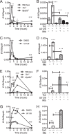
M. tuberculosis induces TLR2- and ERK pathway-dependent gene regulation that appears within 2 h and persists for at least 24 h. Macrophages were pretreated for 1 h with U0126 (10 μM) or DMSO vehicle control, as indicated, and then incubated with or without M. tuberculosis (MOI = 3) for 24 h or the indicated times. RNA was prepared, and gene expression was analyzed by qRT-PCR in triplicate. (A to D) Regulation of Il10. (A and C) Macrophages expressed two phases of Il10, an early peak within 2 h of M. tuberculosis infection and a late elevation at 24 h. Il10 expression in both phases was inhibited by deletion of Tlr2 (A and B), Myd88 (A), or Tpl2 (D) or inhibition of ERK signaling with U0126 (B and C). (E to H) Regulation of Il12b. Expression of Il12b was induced maximally by 8 h of M. tuberculosis infection but declined to near baseline by 24 h. Il12b expression was inhibited by deletion of Tlr2 (E and F) or Myd88 (E). In contrast to Il10, Il12b expression was enhanced when the ERK pathway was blocked by inhibition with U0126 (F and G) or deletion of Tpl2 (H). The data are shown as means ± standard errors of the mean (SEM) and are representative of the results of at least two independent experiments. Statistical significance was determined as described in Materials and Methods. White bars, wild type; grey bars, U0126 treated; black bars, Tlr2−/−; hatched bars, Tpl2−/−. *, wild-type versus Tlr2−/−; +, wild-type versus Myd88−/−; #, DMSO versus U0126; 3 symbols, P < 0.001; 2 symbols, P < 0.01; 1 symbol, P < 0.05.
FIG 3
FIG 3
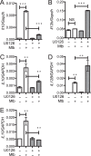
M. tuberculosis regulates cytokine gene transcription in an ERK-dependent manner in murine lung macrophages and human macrophages. Lung macrophages from wild-type mice (A and B), human THP-1 cells (C and D), or human monocyte-derived macrophages (E) were infected with M. tuberculosis (MOI = 3) for 24 h in the continuous presence of U0126 or vehicle control. Total RNA was prepared and analyzed in triplicate by qRT-PCR. The data represent the results of two independent experiments and are graphed as means ± SEM. White bars, DMSO treated; grey bars, U0126 treated. ***, P < 0.001; **, P < 0.01; NS, P > 0.05.
FIG 4
FIG 4
M. tuberculosis-induced ERK signaling promotes IL-10 production and inhibits IL-12 production. Macrophages were incubated with or without U0126 (as for Fig. 2) and infected with M. tuberculosis (MOI = 3) for the indicated times. The supernatants were harvested, and cytokine levels were determined by ELISA. The data shown are representative of the results of three independent experiments and reflect the means ± SEM of biological triplicates handled in parallel throughout the experiment. Statistical comparisons were wild type to Tpl2−/− (*), wild type to Tlr2−/− (+), and wild type untreated to wild type U0126 treated (#); 3 symbols, P < 0.001; 2 symbols, P < 0.01; NS, P > 0.05.
FIG 5
FIG 5
M. tuberculosis-induced TLR2-Tpl2-ERK signaling regulates the balance of macrophage proinflammatory/anti-inflammatory functions, such as Nos2 and arginase 1 expression. Macrophages were treated with U0126 or control treatment, infected with M. tuberculosis for the indicated times (A and B) or for 24 h (C to F), and analyzed for expression of Arg1 and Nos2. (A) Induction of mRNA for markers of anti-inflammatory macrophage states (e.g., Arg1) was dependent on TLR2 and MyD88. Induction of Arg1 was diminished by U0126 or genetic deletion of Tpl2 (B, C, and E), whereas expression of Nos2 (associated with proinflammatory states) was increased by these conditions (D and F). The data are represented as means ± SEM and are representative of at least two independent experiments. Statistical significance was determined as described in Materials and Methods. White bars, wild type; grey bars, U0126 treated; black bars, Tlr2−/−; hatched bars, Tpl2−/−. *, wild type versus Tlr2−/−; +, wild type versus Myd88−/−; #, DMSO versus U0126; 3 symbols, P < 0.001; 2 symbols, P < 0.01; 1 symbol, P < 0.05; NS, P > 0.05.
FIG 6
FIG 6
M. tuberculosis inhibits MHC-II antigen presentation through TLR2-Tpl2-ERK signaling. (A to D) Macrophages were infected with M. tuberculosis for 4 h and washed. IFN-γ (2 ng/ml) was added for an additional 20 h, and RNA was prepared for qRT-PCR analysis. (E) Macrophages were infected for 4 h to allow processing of M. tuberculosis antigen, and naive P25 T cells were then added (1:1 ratio) for 72 h. The T cells were recovered, and Il2 mRNA expression was analyzed by qRT-PCR. (F) Macrophages were infected with M. tuberculosis and cultured with naive P25 T cells for 48 h, and then the supernatants were collected for ELISA measurement of IL-2. (G) Macrophages were cocultured with naive P25 CD4+ T cells and synthetic cognate peptide for 72 h, and the supernatants were analyzed by ELISA for IL-2 secretion. (H) Macrophages were infected with M. tuberculosis for 4 h, washed, treated with IFN-γ for an additional 44 h, stained, and analyzed by flow cytometry to measure the specific median fluorescence intensity (MFI) of MHC-II. The data are means ± SEM and represent the results of at least three independent experiments. White bars, wild type; grey bars, U0126 treated; black bars, Tlr2−/−; hatched bars, Tpl2−/−. ****, P < 0.0001; ***, P < 0.001; **, P < 0.01; NS, P > 0.05.
FIG 7
FIG 7
ERK signaling in macrophages inhibits Th1 polarization in responding T cells. Macrophages were infected with M. tuberculosis and cocultured with P25 T cells. The T cell responses were analyzed as for Fig. 6. (A and B) Expression of the Th1 markers T-bet and IFN-γ is increased following stimulation of naive P25 T cells for 72 h when antigen-presenting macrophages are Tpl2 deficient. (C) Tpl2-deficient macrophages induced higher levels of IFN-γ protein expression by previously primed Th1 effector P25 T cells following 48 h of restimulation. (D) The expression of other T cell-expressed cytokine genes, such as Il4, Il10, Il17a, or Il22, was not substantially altered by knockout of Tpl2 signaling in macrophages following 72 h stimulation of naive P25 T cells. The data represent the results of two independent experiments and show means ± SEM. White bars, wild type; black bars, Tlr2−/−; hatched bars, Tpl2−/−. **, P < 0.01; *, P < 0.05; NS, P > 0.05.
FIG 8
FIG 8
Model of ERK pathway signaling and outcomes in the M. tuberculosis-regulated macrophage. TLR2 signaling represents a major macrophage-intrinsic signaling pathway in response to M. tuberculosis infection, and downstream ERK activation shapes the macrophage response to outcomes that favor establishment of latent M. tuberculosis infection. TLR2-ERK signaling drives macrophages to enhance IL-10 production and diminish IL-12 production. ERK signaling also promotes arginase 1 expression and inhibits expression of MHC-II and iNOS. These phenomena collectively result in reduced presentation of M. tuberculosis antigen to T cells, reduced stimulation of T cells into a Th1 polarization state, and reduced innate macrophage microbicidal mechanisms, such as NO elaboration. If ERK signaling is selectively blocked, M. tuberculosis infection of macrophages results in less expression of IL-10 or arginase 1; higher levels of IL-12, iNOS, and MHC-II; and enhanced Th1 polarization.

References

    1. Cooper AM. 2009. Cell-mediated immune responses in tuberculosis. Annu Rev Immunol 27:393–422. doi:10.1146/annurev.immunol.021908.132703. - DOI - PMC - PubMed
    1. Flynn JL, Chan J. 2001. Immunology of tuberculosis. Annu Rev Immunol 19:93–129. doi:10.1146/annurev.immunol.19.1.93. - DOI - PubMed
    1. Fremond CM, Yeremeev V, Nicolle DM, Jacobs M, Quesniaux VF, Ryffel B. 2004. Fatal Mycobacterium tuberculosis infection despite adaptive immune response in the absence of MyD88. J Clin Invest 114:1790–1799. doi:10.1172/JCI200421027. - DOI - PMC - PubMed
    1. Yahagi A, Umemura M, Tamura T, Kariyone A, Begum MD, Kawakami K, Okamoto Y, Hamada S, Oshiro K, Kohama H, Arakawa T, Ohara N, Takatsu K, Matsuzaki G. 2010. Suppressed induction of mycobacterial antigen-specific Th1-type CD4+ T cells in the lung after pulmonary mycobacterial infection. Int Immunol 22:307–318. doi:10.1093/intimm/dxq010. - DOI - PubMed
    1. Chackerian AA, Perera TV, Behar SM. 2001. Gamma interferon-producing CD4+ T lymphocytes in the lung correlate with resistance to infection with Mycobacterium tuberculosis. Infect Immun 69:2666–2674. doi:10.1128/IAI.69.4.2666-2674.2001. - DOI - PMC - PubMed

Publication types

MeSH terms

LinkOut - more resources