. 2015 Apr 23;125(17):2704-11.
doi: 10.1182/blood-2014-11-611707. Epub 2015 Mar 16.

Merozoite surface protein 1 recognition of host glycophorin A mediates malaria parasite invasion of red blood cells

Affiliations

Merozoite surface protein 1 recognition of host glycophorin A mediates malaria parasite invasion of red blood cells

Michael R Baldwin et al. Blood. .

Abstract

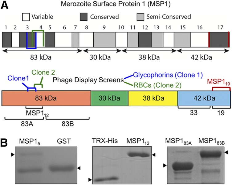
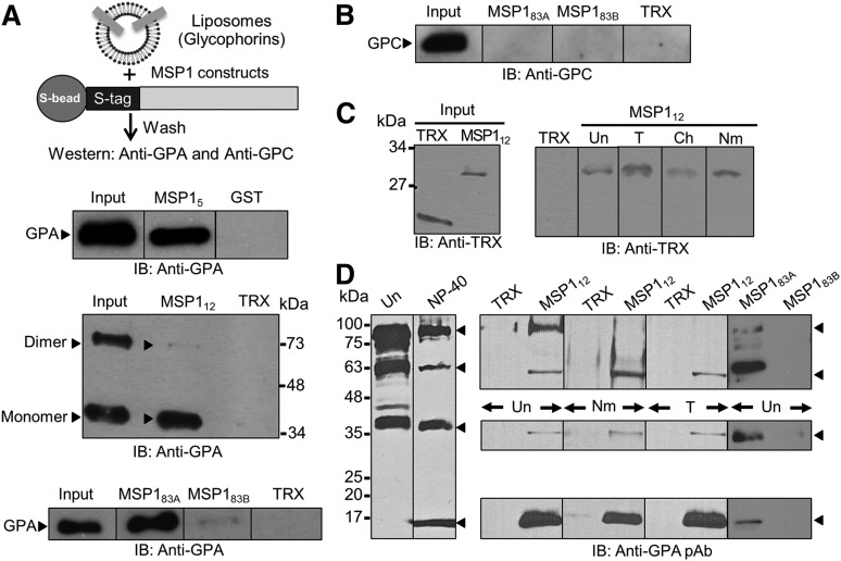
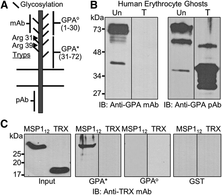
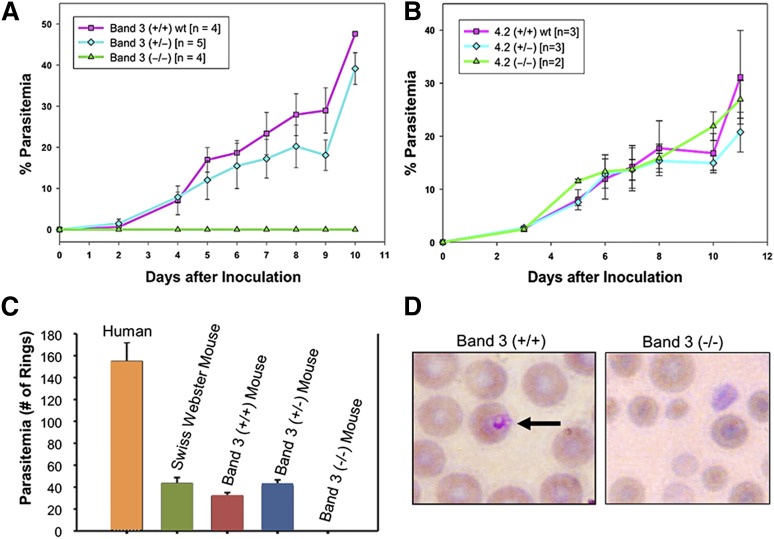
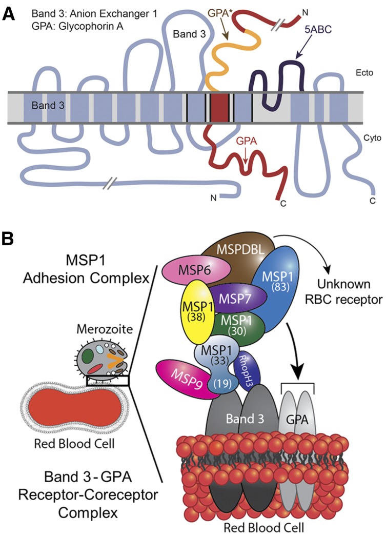
Plasmodium falciparum invasion of human red blood cells (RBCs) is an intricate process requiring a number of distinct ligand-receptor interactions at the merozoite-erythrocyte interface. Merozoite surface protein 1 (MSP1), a highly abundant ligand coating the merozoite surface in all species of malaria parasites, is essential for RBC invasion and considered a leading candidate for inclusion in a multiple-subunit vaccine against malaria. Our previous studies identified an interaction between the carboxyl-terminus of MSP1 and RBC band 3. Here, by employing phage display technology, we report a novel interaction between the amino-terminus of MSP1 and RBC glycophorin A (GPA). Mapping of the binding domains established a direct interaction between malaria MSP1 and human GPA within a region of MSP1 known to potently inhibit P falciparum invasion of human RBCs. Furthermore, a genetically modified mouse model lacking the GPA- band 3 complex in RBCs is completely resistant to malaria infection in vivo. These findings suggest an essential role of the MSP1-GPA-band 3 complex during the initial adhesion phase of malaria parasite invasion of RBCs.


Figures

Figure 1
Figure 1
Domain organization of recombinant MSP1. (A) MSP1 is grouped into 17 blocks classified as conserved, semiconserved, or variable. The diagram shows 4 subunits of MSP1 following the action of subtilisin-1. Two independent overlapping phage clones of MSP1 are indicated. Clone 1, identified in the screen using purified glycophorins, consists of amino acids 204 to 250, whereas clone 2, identified in the screen against neuraminidase-treated RBCs, encodes amino acids 240 to 279. The MSP112 construct incorporates both clones 1 and 2. The respective locations of MSP183A, MSP183B, and MSP119 are also annotated. (B) Coomassie-stained sodium dodecyl sulfate–polyacrylamide gel electrophoresis of MSP1 fusion proteins includes MSP15, containing amino acids 204 to 250 from phage clone 1. MSP112 (amino acids 194-296) expresses a relatively stable protein encompassing both phage clones. MSP183A and MSP183B cover the entire 83-kDa N-terminal subunit of MSP1.
Figure 2
Figure 2
MSP1-GPA binding assays. (A) MSP1 fusion proteins containing an S-tag (GST in MSP15) were immobilized to S-beads (or glutathione beads) and incubated with glycophorin-containing liposomes. Bound proteins were detected by immunoblotting (IB). The TRX fusion protein containing an S-tag and GST were used as negative controls. MSP112 showed specific binding to GPA relative to control in both the monomer (lower band) and dimer species of GPA (upper band). Binding of GPA dimer was detected under mild conditions of beads washings, whereas the GPA monomer binding was observed under both mild and stringent conditions. GPA binding to MSP183A and a very faint signal to MSP183B were also observed. (B) Immunoblotting against GPC did not detect any signal to either MSP183A or MSP183B fusion proteins. (C) RBC binding assay. Bound proteins were eluted by high salt and detected by immunoblotting. MSP112 showed specific binding to RBCs irrespective of their treatment with trypsin (T), chymotrypsin (Ch), or neuraminidase (Nm). Untreated RBCs (Un) were used as control. (D) Detection of GPA species in the detergent-solubilized human RBC ghosts by immunoblotting. Four specific bands of GPA were detected corresponding to homodimer, heterodimer, monomer, and a previously unrecognized 18-kDa band in the NP-40–solubilized ghosts. Immunoblotting using a polyclonal antibody (pAb) against GPA showed specific binding of MSP112 and MSP183A, but not MSP183B, to multiple species of GPA.
Figure 3
Figure 3
Characterization of the MSP1-GPA interaction. (A) GPA, a single transmembrane protein, shows significant glycosylation in the extracellular domain that can be removed by neuraminidase treatment. The approximate locations of the sites recognized by the monoclonal antibody (mAb) and polyclonal antibody (pAb) are indicated. The trypsin cleavage of GPA occurs at 2 distinct arginine residues (Arg31 and Arg39) on the extracellular domain. The extracellular domain of GPA was expressed as 2 nonoverlapping contiguous segments designated as GPAo and GPA*. (B) Both untreated (Un) and trypsin-treated (T) erythrocyte ghosts were tested by immunoblotting (IB) using monoclonal and polyclonal antibodies against GPA. (C) Binding of MSP112 to the trypsin-resistant segment of GPA (GPA*, amino acids 31-72) was detected by immunoblotting against TRX. The trypsin-sensitive segment of GPA (GPAo, amino acids 1-30) was also expressed and tested under identical conditions. Because both fusion proteins contained the GST-tag, GST was also used as a negative control.
Figure 4
Figure 4
Resistance of band 3–GPA–deficient mice to malaria infection. (A) P yoelii 17XL infection to wild-type (band 3+/+), heterozygous (+/−), and band 3–GPA–deficient (−/−) mice in vivo as described in “Materials and methods.” (B) P yoelii challenge in mice deficient of protein 4.2. (C) Human and mouse RBCs were challenged with P falciparum in vitro and the ring-stage parasites were counted. No invasion of P falciparum was detected in band 3–GPA–deficient (−/−) RBCs during the first cycle of invasion in vitro. (D) Representative images of RBCs from infected wild-type (+/+) and band 3–GPA–deficient (−/−) mice (from panel C) indicate the relative intact morphology of mutant RBCs under the in vitro invasion conditions. Wright-Giemsa was used for staining and images were observed at ×100 magnification. It is noteworthy that the putative regions of P falciparum MSP1 that bind to band 3 (MSP119) and GPA (MSP112) show 48.5% and 43.8% sequence identity, respectively, with their counterparts in P yoelii MSP1.
Figure 5
Figure 5
Model of malaria MSP1–RBC receptor complex. (A) Band 3 and GPA are known to exhibit close physical association in the RBC membrane. The 5ABC region of band 3 that binds to malaria MSP119 (dark blue) is adjacent to GPA* (orange), which binds to MSP112, as adapted from Williamson and Toye. (B) Proposed model of malaria merozoite adhesion to erythrocyte by engaging the MSP1 protein complex with host band 3–GPA receptor/coreceptor complex. The recently identified MSPDBL proteins of the MSP1 complex are also shown.

References

    1. Murray CJ, Ortblad KF, Guinovart C, et al. Global, regional, and national incidence and mortality for HIV, tuberculosis, and malaria during 1990-2013: a systematic analysis for the Global Burden of Disease Study 2013. Lancet. 2014;384(9947):1005–1070. - PMC - PubMed
    1. Anders RF, Saul A. Malaria vaccines. Parasitol Today. 2000;16(10):444–447. - PubMed
    1. Delany I, Rappuoli R, De Gregorio E. Vaccines for the 21st century. EMBO Mol Med. 2014;6(6):708–720. - PMC - PubMed
    1. Cowman AF, Berry D, Baum J. The cellular and molecular basis for malaria parasite invasion of the human red blood cell. J Cell Biol. 2012;198(6):961–971. - PMC - PubMed
    1. Farrow RE, Green J, Katsimitsoulia Z, Taylor WR, Holder AA, Molloy JE. The mechanism of erythrocyte invasion by the malarial parasite, Plasmodium falciparum. Semin Cell Dev Biol. 2011;22(9):953–960. - PubMed

Publication types

MeSH terms