. 2015 May:112:139-50.
doi: 10.1016/j.biochi.2015.03.006. Epub 2015 Mar 13.

Inhibitor of differentiation 4 (ID4) acts as an inhibitor of ID-1, -2 and -3 and promotes basic helix loop helix (bHLH) E47 DNA binding and transcriptional activity

Affiliations

Inhibitor of differentiation 4 (ID4) acts as an inhibitor of ID-1, -2 and -3 and promotes basic helix loop helix (bHLH) E47 DNA binding and transcriptional activity

Pankaj Sharma et al. Biochimie. 2015 May.

Abstract

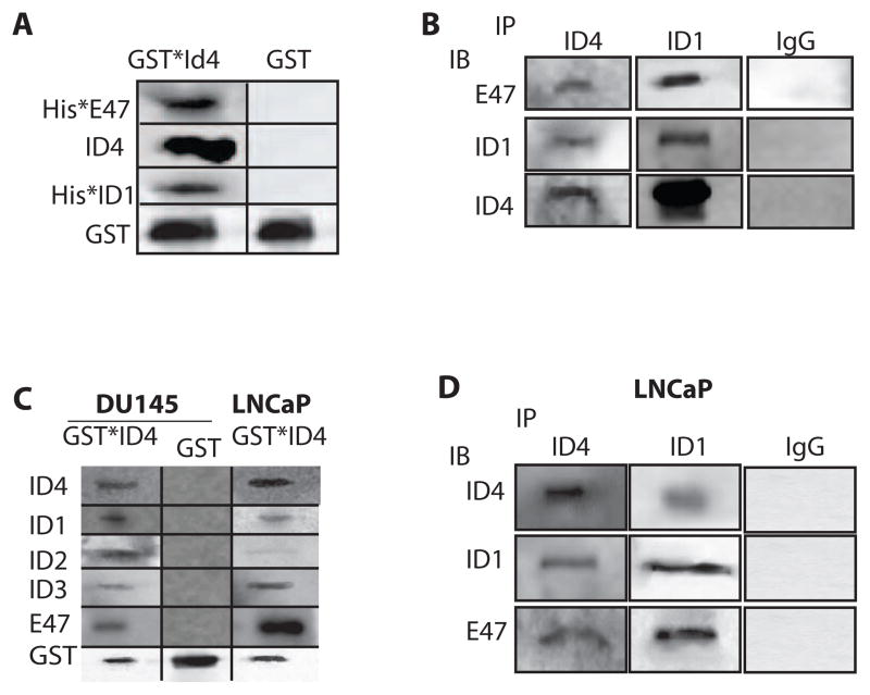
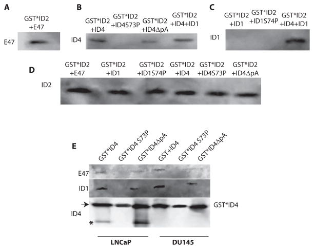
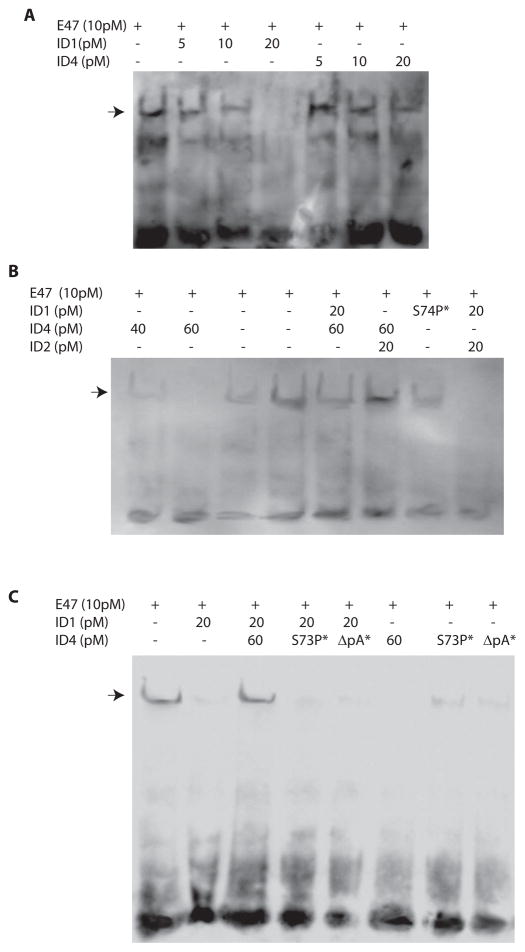
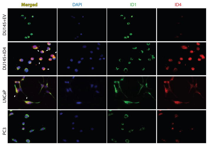
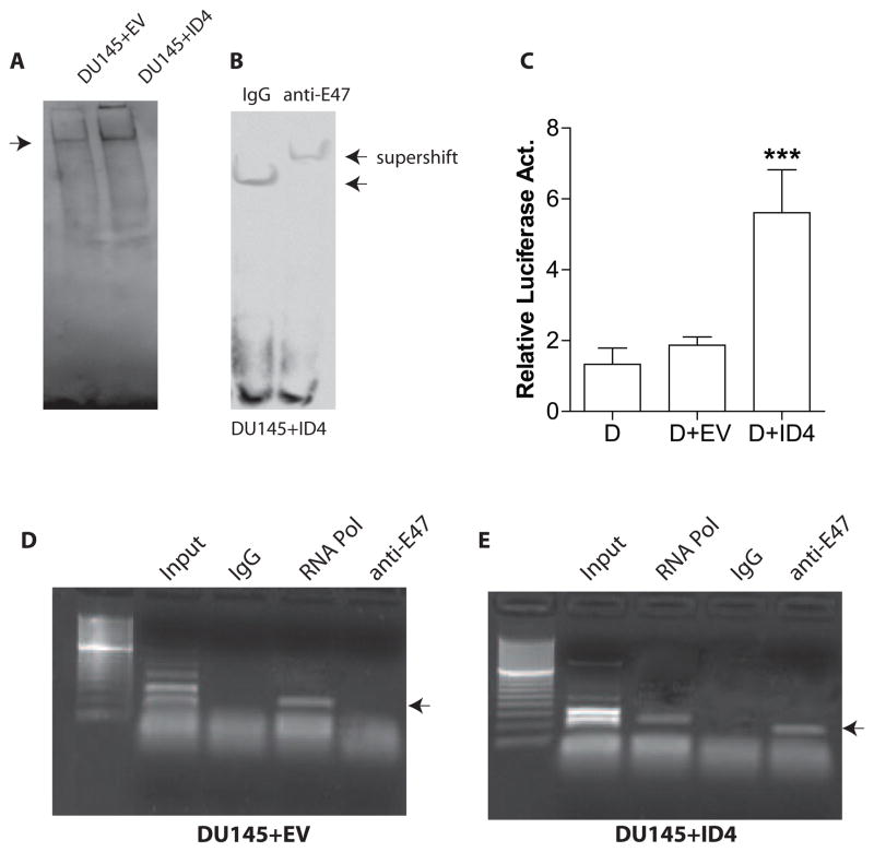
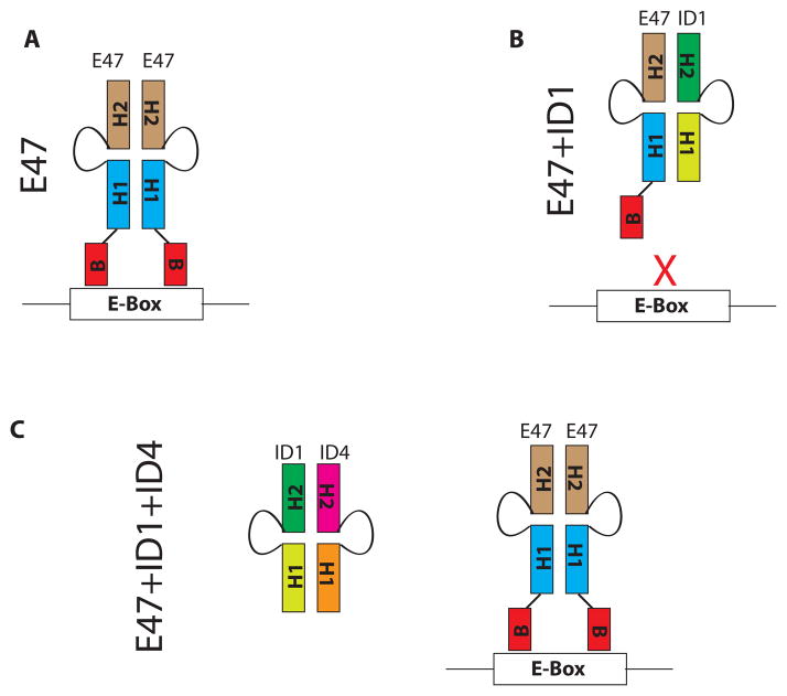
The four known ID proteins (ID1-4, Inhibitor of Differentiation) share a homologous helix loop helix (HLH) domain and act as dominant negative regulators of basic-HLH transcription factors. ID proteins also interact with many non-bHLH proteins in complex networks. The expression of ID proteins is increasingly observed in many cancers. Whereas ID-1, ID-2 and ID-3, are generally considered as tumor promoters, ID4 on the contrary has emerged as a tumor suppressor. In this study we demonstrate that ID4 heterodimerizes with ID-1, -2 and -3 and promote bHLH DNA binding, essentially acting as an inhibitor of inhibitors of differentiation proteins. Interaction of ID4 was observed with ID1, ID2 and ID3 that was dependent on intact HLH domain of ID4. Interaction with bHLH protein E47 required almost 3 fold higher concentration of ID4 as compared to ID1. Furthermore, inhibition of E47 DNA binding by ID1 was restored by ID4 in an EMSA binding assay. ID4 and ID1 were also colocalized in prostate cancer cell line LNCaP. The alpha helix forming alanine stretch N-terminal, unique to HLH ID4 domain was required for optimum interaction. Ectopic expression of ID4 in DU145 prostate cancer line promoted E47 dependent expression of CDKNI p21. Thus counteracting the biological activities of ID-1, -2 and -3 by forming inactive heterodimers appears to be a novel mechanism of action of ID4. These results could have far reaching consequences in developing strategies to target ID proteins for cancer therapy and understanding biologically relevant ID-interactions.

Keywords: Cancer; DNA-Protein interaction; ID4; Protein–protein interaction; Tumor suppressor gene.


Conflict of interest statement

Conflict of Interest: The authors declare no conflict of interest

Figures

Figure 1
Figure 1
Sequence alignments and phylogenetic analysis of ID proteins. A: Sequence alignments of full length ID-1, -2, -3 and -4. The conserved residues are highlighted. The Helix1, loop and Helix II of the conserved HLH (Helix-loop-Helix) domain is shown by double sided arrows. The alanine stretch in ID4 that was deleted by site directed mutagenesis is indicated by ΔpAla (deletion of poly Alanine tract). The asterisk indicates the position of conserved serine in helix I which was mutated to proline in ID1 (S74P) and ID4 (S73P). B: The phylogenetic analysis of ID-1, -2, -3 and -4 based on structural homology. C: The alignment of ID1, ID2, ID3 and ID4 HLH domain only. D: Phylogenetic relationship between ID-1, -2, -3 and -4 based on HLH only domain homology. The sequence alignments and phylogenetic analysis were performed using MAFFT (Multiple Alignment using Fast Fourier Transform).
Figure 2
Figure 2
Interaction of ID4 with ID-1, -2 and -3. A: On column interaction of Glutathione S-transferase coupled ID4 (GST*ID4) with recombinant Histidine (6x His) tagged E47 (His*E47), His*ID1. The proteins were eluted with 5mM glutathione and the elute was subjected to immuno-blot analysis with protein specific antibodies. The GST tag alone was used as a negative control. B: The antibodies against ID4 or ID1 immobilized on protein A Mag beads were incubated with recombinant ID4, His*ID1 and His*E47. The elute was subjected to immuno-blot (IB) analysis with protein specific antibodies. Non-immune rabbit IgG immobilized on protein A Mag beads and incubated with recombinant proteins as above was used as negative control. C: On column interaction of total cellular proteins isolated from LNCaP and DU145 prostate cancer cell lines with GST*ID4. The bound proteins were eluted with glutathione and subjected to immuno blot analysis with specific antibodies. D: In a format similar to panel B above, the ID4 and ID1 antibodies immobilized on protein A Mag beads were incubated with total cellular proteins from LNCaP cells (immunoprecipitation, IP) followed by elution and IB. The data is representative of at least 4 experiments.
Figure 3
Figure 3
Interaction of wild type ID4 and its various mutants with recombinant ID2 and cellular E47 and ID1. All GST based interactions were on the GST affinity column. The bound proteins were eluted as a complex with thrombin (when GST*ID2 was used as bait, panels A, B, C and D) or glutathione (Panel E) and subjected to immuno-blot using protein specific antibodies indicated on the left side of the blot. A: Interaction between GST*ID2 and E47 was used as a positive control. B, C and D: Interaction of GST*ID2 with ID4, ID4S73P (ID4 HLH mutant) or ID4ΔpA (ID4 in which the alanine tract was deleted, see Fig. 1). The elute was probed with ID4 antibody (panel B), ID1 antibody (Panel C) or ID2 antibody (Panel D, as loading control). E: Interaction of GST*ID4, GST*ID4S73P and GST*ID4ΔpA with total cellular proteins from LNCaP and DU145 cells. The elute was probed with E47, ID1 and ID4 antibodies. The asterisk in the ID4 immuno-blot represents to location of native, cellular ID4 as opposed to GST-ID4 marked by an arrow. Representative of at least 4 experiments is shown.
Figure 4
Figure 4
Electrophoretic mobility shift assay (EMSA) demonstrating interaction between double stranded consensus E-Box oligonucleotide and recombinant E47 in the presence or absence of recombinant ID1 and/or ID4. A, B and C: The double stranded E-Box oligonucleotide was incubated with 10pM of recombinant E47 either alone or in the presence of ID1 (A, B and C), ID4 (A, B and C), ID2 (B), HLH mutant (S74P) of ID1 (B), HLH mutant of ID4 (S73P, C) or with ID4 in which alanine tract was deleted ΔpA, C). The GST tag from ID4 (and its mutants) and ID2 was removed with respective on column protease digestion. The E47 and ID1 retained the histidine tag. All concentrations are in Pico moles (pM). The ID1 S75P* and ID4 (S73P* and ΔpA*) mutants were used at 20pM and 60pM respectively. The arrows indicate the gel shift due to the binding between E-Box oligonucleotide and E47. The free probe is at the bottom of the gel. The data is representative of at least 3 different experiments using recombinant proteins from different batches.
Figure 5
Figure 5
ColocalizationColocalization of ID1 and ID4 in prostate cancer cell lines LNCaP, DU145 and PC3. ID1 (green) and ID4 (red) were detected by fluorescence based immunocytochemistry. The merged images were used to detect colocalization (yellow, Merged) of ID1 and ID4. The nuclei were stained with DAPI. Immuno-localization studies were also performed in DU145 cells in which ID4 was over expressed (DU145+ID4) and compared to cells transfected with vector alone (DU145+EV). The arrows indicate colocalization which was observed in LNCaP and DU145+ID4 cells. No colocalization was observed in DU145+EV cells due to the lack of expression of ID4. A representative image of three different experiments is shown. All images are at 200X magnification.
Figure 6
Figure 6
E-Box binding and transcriptional activity in DU145 cells over-expressing ID4. A: Four microgram (4ug) of nuclear extracts from DU145+EV (empty vector) and DU145+ID4 cells were used in the EMSA to demonstrate relative binding to consensus E-Box. Though not quantitative, a stronger signal of the shifted band (indicated by arrow) in DU145+ID4 suggests higher E-Box DNA binding relative to DU145+EV cells. B: The supershift using E47 specific antibody was to ascertain that the gel shift using DU145+ID4 nuclear extracts was due to the binding of E47 to E-Box oligonucleotide. Non-immune IgG was used as a negative that did not result in a supershift. C: Relative E-Box luciferase reporter activity in DU145 (D), DU145+EV (D+EV) and DU145+ID4 (D+ID4) cells. The results were first normalized to Renilla luciferase and then to DU145 cells (set to 1). The data is expressed as mean+SEM of three experiments in triplicate. The significance of differences between means were analyzed by student’s t-test (***: P<0.001). D and E: Chromatin immunoprecipitation (ChIP) of E47 on CDKN1A proximal promoter in DU145+EV (D) and DU145+ID4 (E) cells. A clear PCR band (anti-E47 lane) was observed on the reverse cross-linked product in DU145+ID4 cells using primers around the E-Box site known to bind E47. A similar product was absent/un-detectable in DU145+EV cells. The lanes with Input, IgG alone (negative control) and RNA Pol II is shown. The RNA PolII was observed in DU145+ID4 cells suggesting active transcription of CDKN1A. Representative data of three different experiments is shown.
Figure 7
Figure 7
Proposed schematic representing the mechanism by which ID4 restores bHLH-DNA interaction. A: bHLH proteins such as E47 bind DNA as homodimers or heterodimers with tissue bHLH proteins. The dimerization involves the conserved helix-loop-helix domain (represented by H1 and H2 with intertwining loop) whereas the basic domain (B) binds the E-Box. B: ID1 heterodimerizes with bHLH proteins through the conserved HLH domain. The lack of basic domain in ID1 renders the heterodimer incapable of binding the DNA. Thus ID1 essentially acts as a dominant negative inhibitor of bHLH transcription factors. C: ID4, a distant member of the ID protein family could acts as an inhibitor of inhibitor of differentiation proteins such as ID1. ID4 hetero-dimerizes with ID1 which allows bHLH dimerization, subsequent DNA binding and transcriptional regulation of E-Box dependent genes.

Similar articles

Cited by

References

    1. Massari ME, Murre C. Helix-loop-helix proteins: regulators of transcription in eucaryotic organisms. Mol Cell Biol. 2000;20(2):429–440. - PMC - PubMed
    1. Ledent V, Vervoort M. The basic helix-loop-helix protein family: comparative genomics and phylogenetic analysis. Genome Res. 2001;11(5):754–770. - PMC - PubMed
    1. Atchley WR, Fitch WM. A natural classification of the basic helix-loop-helix class of transcription factors. Proc Natl Acad Sci U S A. 1997;94(10):5172–5176. - PMC - PubMed
    1. Murre C, Bain G, van Dijk MA, Engel I, Furnari BA, Massari ME, Matthews JR, Quong MW, Rivera RR, Stuiver MH. Structure and function of helix-loop-helix proteins. Biochim Biophys Acta. 1994;1218(2):129–135. - PubMed
    1. Murre C, McCaw PS, Vaessin H, Caudy M, Jan LY, Jan YN, Cabrera CV, Buskin JN, Hauschka SD, Lassar AB, Weintraub H, Baltimore D. Interactions between heterologous helix-loop-helix proteins generate complexes that bind specifically to a common DNA sequence. Cell. 1989;58(3):537–544. - PubMed

Publication types

MeSH terms

Substances