

[Classification of amyloidosis by laser micro-dissection and mass spectrometry based proteomic analysis]
- PMID: 25778882
- PMCID: PMC7342159
- DOI: 10.3760/cma.j.issn.0253-2727.2015.02.003
[Classification of amyloidosis by laser micro-dissection and mass spectrometry based proteomic analysis]
Abstract
Objective: To establish a novel method to determine specific type of amyloidosis through laser microdissection and mass spectrometry (LMD/MS) based proteomic analysis.
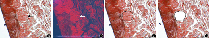
Methods: There were 138 formalin-fixed and paraffin-embedded (FFPE) biopsy samples of patients who were diagnosed as systemic amyloidosis used in this study. For each case, a 10 μm section stained with congo-red and positive amyloid deposits were identified under fluorescent light, followed by micro-dissection and mass spectrometry analysis. The amyloidosis subtype was confirmed based on the most abundant amyloid protein.
Results: The tissue types of 138 specimens were as following: subcutaneous abdominal fat accounted for 26%, tongue for 19%, gingiva for 11%, kidney for 9%, intestine for 9%, heart for 6% and others for 20%. Specific types of amyloid were accurately detected in 121 cases, including 106 (87.6%) amyloid light chain (AL) type, 7 (5.8%) amyloid trans-thy-retin (ATTR), 2 (1.7%) amyloidogenic protein A (AA), 2 (1.7%) amyloid heavy chain (AH)/AL+AH, 2 (1.7%) fibrinogen alpha chain (AFib), 1(0.8%) amyloid apolipoprotein A-type II (AApoA-II) and one (0.8%) amyloid lysozyme (ALys). Diagnosis of amyloidosis was excluded in 5 cases. The types of twelve cases were indeterminate by LMD/MS. On the whole, LMD/MS reached 91.3% accuracy rate in amyloid typing. Commonly involved organs (for example, heart, kidney and liver) turned out to be suitable sources of FFPE samples with typing success rate of almost 100%. In contrast, MS analysis was successful in only 83.3% of subcutaneous abdominal fat samples.
Conclusion: LMD/MS method provided a more direct technique for accurate typing of amyloidosis in a single procedure.
目的: 评估利用激光显微切割联合质谱蛋白质组学技术对系统性淀粉样变性进行分型的有效性。
方法: 以138例病理确诊为淀粉样变性患者的经福尔马林固定石蜡包埋标本为研究对象。利用显微切割收集刚果红染色阳性区域组织并行质谱蛋白质组学分析,以指数修正的蛋白质丰度指标(emPAI)为标准,根据丰度最高的致淀粉样变蛋白判定淀粉样变性亚型。
结果: 在138份组织标本中,腹壁脂肪占26%,舌体占19%,齿龈占11%,肾脏占9%,胃肠道占9%,心脏占6%,其余类型样本占20%。总共121例患者获得成功分型,包括轻链型106例(87.6%),遗传性甲状腺转运球蛋白型7例(5.8%),致淀粉样变蛋白A型、重链/重链+轻链型及纤维蛋白原α链型各2例(1.7%),载脂蛋白A-Ⅱ型及溶菌酶型各1例(0.8%)。5例被排除淀粉样变性诊断,12例分型失败。总体诊断准确率为91.3%。在各种组织类型标本中,腹壁脂肪组织的分型成功率最低,仅为83.3%。
结论: 激光显微切割联合质谱蛋白质组学方法能有效鉴定出淀粉样变性亚型。
Figures
Similar articles
-
A stepwise data interpretation process for renal amyloidosis typing by LMD-MS.BMC Nephrol. 2022 Apr 13;23(1):144. doi: 10.1186/s12882-022-02785-9. BMC Nephrol. 2022. PMID: 35418036 Free PMC article.
-
Mass spectrometry-based proteomic diagnosis of renal immunoglobulin heavy chain amyloidosis.Clin J Am Soc Nephrol. 2010 Dec;5(12):2180-7. doi: 10.2215/CJN.02890310. Epub 2010 Sep 28. Clin J Am Soc Nephrol. 2010. PMID: 20876678 Free PMC article.
-
Mass Spectrometry Amyloid Typing Is Reproducible across Multiple Organ Sites.Biomed Res Int. 2019 Jan 31;2019:3689091. doi: 10.1155/2019/3689091. eCollection 2019. Biomed Res Int. 2019. PMID: 30834260 Free PMC article.
-
[New advances in the subtyping of systemic amyloidosis].Zhongguo Shi Yan Xue Ye Xue Za Zhi. 2014 Feb;22(1):259-62. doi: 10.7534/j.issn.1009-2137.2014.01.052. Zhongguo Shi Yan Xue Ye Xue Za Zhi. 2014. PMID: 24598691 Review. Chinese.
-
Pathology and diagnosis of renal non-AL amyloidosis.J Nephrol. 2018 Jun;31(3):343-350. doi: 10.1007/s40620-017-0426-6. Epub 2017 Aug 21. J Nephrol. 2018. PMID: 28828707 Review.
Cited by
-
Diagnosis for Chinese patients with light chain amyloidosis: a scoping review.Ann Med. 2023 Dec;55(1):2227425. doi: 10.1080/07853890.2023.2227425. Ann Med. 2023. PMID: 37387123 Free PMC article.
-
Prognostic Value of Holter Monitoring in Light Chain Amyloidosis.J Clin Med. 2023 Dec 1;12(23):7457. doi: 10.3390/jcm12237457. J Clin Med. 2023. PMID: 38068514 Free PMC article.
References
-
- Merlini G, Bellotti V. Molecular mechanisms of amyloidosis[J] N Engl J Med. 2003;349(6):583–596. - PubMed
-
- Sipe JD, Benson MD, Buxbaum JN, et al. Amyloid fibril protein nomenclature: 2010 recommendations from the nomenclature committee of the International Society of Amyloidosis[J] Amyloid. 2010;17(3-4):101–104. - PubMed
-
- Vrana JA, Gamez JD, Madden BJ, et al. Classification of amyloidosis by laser microdissection and mass spectrometry-based proteomic analysis in clinical biopsy specimens[J] Blood. 2009;114(24):4957–4959. - PubMed
MeSH terms
Substances
LinkOut - more resources
Full Text Sources
Medical
Research Materials