

Comment
doi: 10.1083/jcb.201501107.
Balancing cell behavior at boundaries
Affiliations
- PMID: 25778916
- PMCID: PMC4362469
- DOI: 10.1083/jcb.201501107
Item in Clipboard
Comment
Balancing cell behavior at boundaries
J Cell Biol.
.
Abstract
The restriction of cell intermingling across boundaries is essential for the establishment of discrete tissues. Eph receptor signaling prevents intermingling at many boundaries. In this issue, Luu et al. (2015. J. Cell Biol. http://dx.doi.org/10.1083/jcb.201409026) report a parallel pathway, mediated by Wnt signaling, Snail1, and paraxial protocadherin (PAPC). This pathway establishes a distinctive organization of cell adhesion and intercellular gaps at the interface between tissues.
© 2015 Wilkinson.
Figures
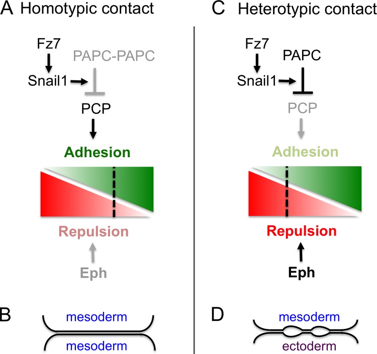
Signaling and responses to cell interactions. (A) At homotypic contacts of mesoderm cells, the PCP pathway can promote adhesion (green) because PAPC complexes form that have low PCP inhibitory activity. Eph receptor activation that promotes repulsion (red) is weak because coexpressed ephrins have low affinity. Consequently, the balance of cell responses favors adhesion (B). (C) At heterotypic contacts, free PAPC inhibits the PCP pathway. This PAPC activity requires Fz7-induced expression of Snail1. Eph receptors are strongly activated by high affinity ephrins expressed in ectoderm. Consequently, there is a balance of repulsion and adhesion that leads to formation of cleft contacts, characterized by interspersed stretches of adhesion and intercellular gaps (D).
Comment on
-
PAPC mediates self/non-self-distinction during Snail1-dependent tissue separation.J Cell Biol. 2015 Mar 16;208(6):839-56. doi: 10.1083/jcb.201409026. J Cell Biol. 2015. PMID: 25778923 Free PMC article.
References
Publication types
MeSH terms
Substances
Grants and funding
LinkOut - more resources
Full Text Sources
Other Literature Sources
Research Materials
Miscellaneous
