. 2015 Mar 16;208(6):671-81.
doi: 10.1083/jcb.201410047.

The nucleoporin gp210/Nup210 controls muscle differentiation by regulating nuclear envelope/ER homeostasis

Affiliations

The nucleoporin gp210/Nup210 controls muscle differentiation by regulating nuclear envelope/ER homeostasis

J Sebastian Gomez-Cavazos et al. J Cell Biol. .

Abstract

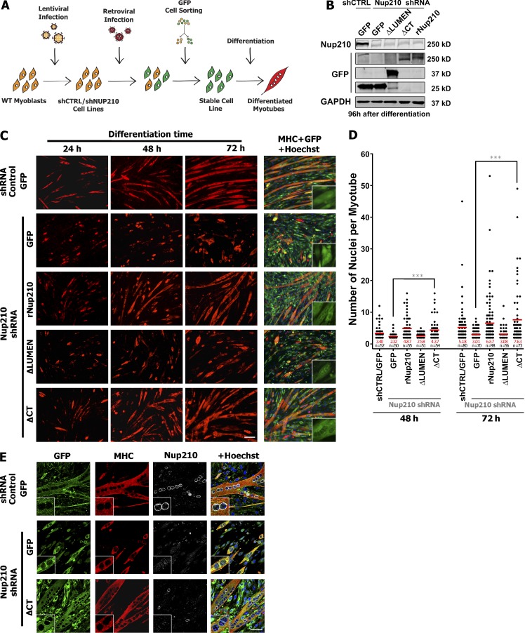
Previously, we identified the nucleoporin gp210/Nup210 as a critical regulator of muscle and neuronal differentiation, but how this nucleoporin exerts its function and whether it modulates nuclear pore complex (NPC) activity remain unknown. Here, we show that gp210/Nup210 mediates muscle cell differentiation in vitro via its conserved N-terminal domain that extends into the perinuclear space. Removal of the C-terminal domain, which partially mislocalizes gp210/Nup210 away from NPCs, efficiently rescues the differentiation defect caused by the knockdown of endogenous gp210/Nup210. Unexpectedly, a gp210/Nup210 mutant lacking the NPC-targeting transmembrane and C-terminal domains is sufficient for C2C12 myoblast differentiation. We demonstrate that the endoplasmic reticulum (ER) stress-specific caspase cascade is exacerbated during Nup210 depletion and that blocking ER stress-mediated apoptosis rescues differentiation of Nup210-deficient cells. Our results suggest that the role of gp210/Nup210 in cell differentiation is mediated by its large luminal domain, which can act independently of NPC association and appears to play a pivotal role in the maintenance of nuclear envelope/ER homeostasis.


Figures

Figure 1.
Figure 1.
Overexpression of Nup210 lacking its C-terminal domain accelerates myotube formation in C2C12 cells. (A) Schematic representation of the topology of Nup210. S.S., signal sequence; TM, transmembrane domain; CT, C-terminal domain. (B) Conservation analysis of Nup210 domains across metazoans: H. sapiens, M. musculus, D. rerio, S. purpuratus, D. melanogaster, and C. elegans. 10 = conserved; 0 = not conserved. Red bars highlight residues that are 100% conserved across all metazoans analyzed. N, N terminus; C, C terminus. (C) Schematic representation of Nup210 GFP fusion constructs overexpressed in C2C12 cells. (D) C2C12 myoblasts were infected with retrovirus carrying GFP alone, Nup210-GFP, Nup210ΔLUMEN-GFP, and Nup210ΔCT-GFP and induced to differentiate. Immunofluorescence against MHC (red) and GFP (green) was performed at 48, 72, and 96 h after differentiation. Hoechst (blue) was used as a nuclear stain. Insets show a digital magnification (3×) of the GFP channel. Bar, 100 µm. (E) Quantification of nuclei in MHC-positive cells (two or more nuclei) at 48, 72, and 96 h after differentiation. Data were collected from three independent experiments and plotted. *, P < 0.05; and **, P < 0.01 indicate a significant difference between overexpression experiments carrying GFP and Nup210ΔCT-GFP by t test. Red bars and numbers represent means.
Figure 2.
Figure 2.
Nup210ΔCT is sufficient to restore C2C12 differentiation. (A) Schematic representation of rescue experiments methodology. C2C12 myoblasts were first infected with lentivirus carrying control and Nup210 shRNAs followed by retroviral transduction of GFP or Nup210 constructs. Cells were then GFP sorted to obtain high expressors. (B) Protein levels of endogenous Nup210 and GFP-tagged Nup210 fragments in reconstituted cell lines expressing scrambled or Nup210 shRNAs. GAPDH was used a loading control. shCTRL, control shRNA. (C) C2C12 myoblasts expressing scrambled or Nup210 shRNAs were infected with retrovirus carrying GFP alone or shRNA-resistant Nup210-GFP, Nup210ΔLUMEN-GFP, and Nup210ΔCT-GFP and induced to differentiate. Immunofluorescence against MHC (red) and GFP (green) was performed at 24, 48, and 72 h after differentiation. Insets show a digital magnification (3×) of GFP channel. Bar, 100 µm. (D) Quantification of nuclei in MHC-positive cells (two or more nuclei) at 48 and 72 h after differentiation from experiments shown in C. Data were collected from four independent experiments. ***, P < 0.001 indicates a significant difference between rescue experiments of GFP and Nup210ΔCT-GFP by t test. Red bars and numbers represent means. (E) Immunofluorescence against GFP (green), MHC (red), and Nup210 (C-terminal–specific antibody, white) in C2C12 cells carrying control or Nup210 shRNAs reconstituted with GFP or Nup210ΔCT-GFP 96 h after differentiation. Hoechst (blue) was used as a nuclear stain. Insets show a digital magnification (3×) of each respective channel. Bar, 50 µm.
Figure 3.
Figure 3.
Nup210ΔCT truncation mutant fails to accumulate at the NE and is partially mislocalized from NPCs. (A) NE distribution of Sec61-β–GFP, NDC1-GFP, Nup210-GFP, Nup210ΔLUMEN-GFP, or Nup210ΔCT-GFP in stable myoblast cell lines. C2C12 cells were stained for GFP (green) and nuclear pores using mAb414 (red). Experiments were performed at least three times independently. Representative data for each condition is shown. Bar, 5 µm. (B) GFP and mAb414 signal profiles at NE cross sections were determined by ImageJ. (C) Localization of GFP and mAb414 signals at NE surfaces of C2C12 myoblasts stably expressing Sec61-β–GFP (n = 555), NDC1-GFP (n = 603), Nup210-GFP (n = 510), Nup210ΔLUMEN-GFP (n = 502), and Nup210ΔCT-GFP (n = 652) where n represents the number of NPCs quantified. C2C12 cells were stained for GFP and nuclear pores using mAb414. Experiments were repeated three times, and representative images are presented. Bar, 1 µm. (D) GFP and mAb414 signal profiles at the NE surface were determined by ImageJ. Colocalization percentage of nuclear pore signals with GFP signals was determined using Imaris.
Figure 4.
Figure 4.
Nup210 mutant lacking NPC sorting signals rescues myotube formation. (A) Schematic representation of Nup210ΔCTLap2-βTM-GFP. N, N terminus; C, C terminus. (B) NE distribution of Nup210ΔCTLap2-βTM-GFP in myoblasts. C2C12 cells were stained for GFP and nuclear pores using mAb414. Bar, 5 µm. (C) Structured illumination microscopy in NE surfaces of C2C12 myoblasts stably expressing Nup210ΔCTLap2-βTM-GFP. Cells were stained for GFP and nuclear pores using mAb414. Bar, 1 µm. (D) Localization of GFP (green line) and mAb414 (red line) signals at the NE of Nup210ΔCTLap2-βTM-GFP (n = 547) was determined by using ImageJ. Colocalization percentage of nuclear pore signals with GFP signals was determined using Imaris. (E) C2C12 myoblasts expressing scrambled or Nup210 shRNAs were infected with retrovirus carrying GFP alone or shRNA-resistant Nup210ΔCTLap2-βTM-GFP and induced to differentiate. Immunofluorescence against MHC (red) and GFP (green) was performed at 24, 48, and 72 h after differentiation. Insets show a digital magnification (3×) of GFP channel. Bar, 100 µm. (F) Quantification of MHC-positive cells with two or more nuclei at 48 and 72 h after differentiation from experiments shown in E. Data were collected from three independent experiments. ***, P < 0.001; and **, P < 0.01 indicate a significant difference between rescue experiments of GFP and Nup210ΔCTLap2-βTM-GFP by t test. Red bars and numbers represent means. (G) Immunofluorescence against GFP (green), MHC (red), and Nup210 (C-terminal–specific antibody, white) in C2C12 cells carrying control or Nup210 shRNAs reconstituted with GFP or Nup210ΔCTLap2-βTM-GFP 96 h after differentiation. Hoechst (blue) was used as a nuclear stain. Insets show a digital magnification (3×) of each respective channel. Bar, 50 µm. (H) FRAP analysis in NE membranes of C2C12 myoblasts stably expressing Nup210ΔCTLap2-βTM-GFP (t1/2 = 64.64 ± 7.10 s; MFR = 0.715 ± 0.019; n = 10), Nup210-GFP (t1/2 = 108.5 ± 12.61 s; MFR = 0.476 ± 0.027; n = 25), and Sec61-β–GFP (t1/2 = 44.8 ± 2.48; MFR = 44.8 ± 2.48; n = 16). White boxes represent photobleached areas. Error bars represent standard deviation of the mean. Bar, 10 µm.
Figure 5.
Figure 5.
Inhibition of C2C12 differentiation by Nup210 depletion leads to an ER stress profile and can be rescued by ER stress inhibitors. (A and B) Western blot analysis in attached and detached cells from Nup210 knockdown cultures 48 h after differentiation, showing scramble shRNA attached cells (negative control) and attached plus detached cells treated with 500 ng/ml tunicamycin (24 h, positive control). (A) Blot of caspase-12, -9, and -3 activation. (B) Blot of ATF6 activation, BiP, and PDI. (C) PDI immunofluorescence in differentiated cultures (attached + detached) 48 h after differentiation expressing scrambled or Nup210 shRNAs. Hoechst was used to stain nuclei. Images were taken at the confocal plane of detached cells. Insets show a digital magnification (3×) of red and blue channels. (D) Differentiation of myoblasts expressing control or Nup210 shRNAs in the presence of TUDCA (500 µg/ml) or vehicle (doubled-distilled H2O). Immunofluorescence against MHC (red) was performed at 24, 48, and 72 h after differentiation. (E) Quantification of nuclei in MHC-positive cells (two or more nuclei) at 24, 48, and 72 h after differentiation from experiments shown in D. Data were collected from three independent experiments. ***, P < 0.001 indicates a significant difference between Nup210 knockdown cells treated with TUDCA or vehicle by t test. Red bars and numbers represent means. (F) Immunofluorescence against Cleaved caspase-3 (green) on differentiated myoblasts expressing control (n = 6,086) or Nup210 shRNAs in the presence of TUDCA (500 µg/ml; n = 4,402), or vehicle (double-distilled H2O; n = 4,612) 72 h after differentiation. Percentages indicate the proportion of cells positive for Cleaved caspase-3 staining versus total number of cells (n). (G) Differentiation of C2C12 cells carrying Nup210 shRNAs expressing GFP alone or MAGE-A3–GFP. Immunofluorescence against MHC (red) and GFP (green) was performed at 24, 48, and 72 h after differentiation. (H) Quantification of nuclei in MHC-positive cells (two or more nuclei) at 48 and 72 h after differentiation from experiments shown in G. Data were collected from three independent experiments. ***, P < 0.001 indicates a significant difference between rescue experiments of GFP and MAGE-A3–GFP by t test. Red bars and numbers represent means. shCtrl, control shRNA. Bars: (C) 50 µm; (D, F, and G) 100 µm.

References

    1. Akhtar A., and Gasser S.M.. 2007. The nuclear envelope and transcriptional control. Nat. Rev. Genet. 8:507–517 10.1038/nrg2122 - DOI - PubMed
    1. Allen N.P., Huang L., Burlingame A., and Rexach M.. 2001. Proteomic analysis of nucleoporin interacting proteins. J. Biol. Chem. 276:29268–29274 10.1074/jbc.M102629200 - DOI - PubMed
    1. Alter J., and Bengal E.. 2011. Stress-induced C/EBP homology protein (CHOP) represses MyoD transcription to delay myoblast differentiation. PLoS ONE. 6:e29498 10.1371/journal.pone.0029498 - DOI - PMC - PubMed
    1. Amaral J.D., Viana R.J., Ramalho R.M., Steer C.J., and Rodrigues C.M.. 2009. Bile acids: regulation of apoptosis by ursodeoxycholic acid. J. Lipid Res. 50:1721–1734 10.1194/jlr.R900011-JLR200 - DOI - PMC - PubMed
    1. Brown C.R., and Silver P.A.. 2007. Transcriptional regulation at the nuclear pore complex. Curr. Opin. Genet. Dev. 17:100–106 10.1016/j.gde.2007.02.005 - DOI - PubMed

Publication types

LinkOut - more resources