Review
. 2015 Apr;56(4):505-13.
doi: 10.1111/epi.12939. Epub 2015 Feb 28.

WONOEP appraisal: molecular and cellular imaging in epilepsy

Affiliations
Review

WONOEP appraisal: molecular and cellular imaging in epilepsy

Kyle P Lillis et al. Epilepsia. 2015 Apr.

Abstract

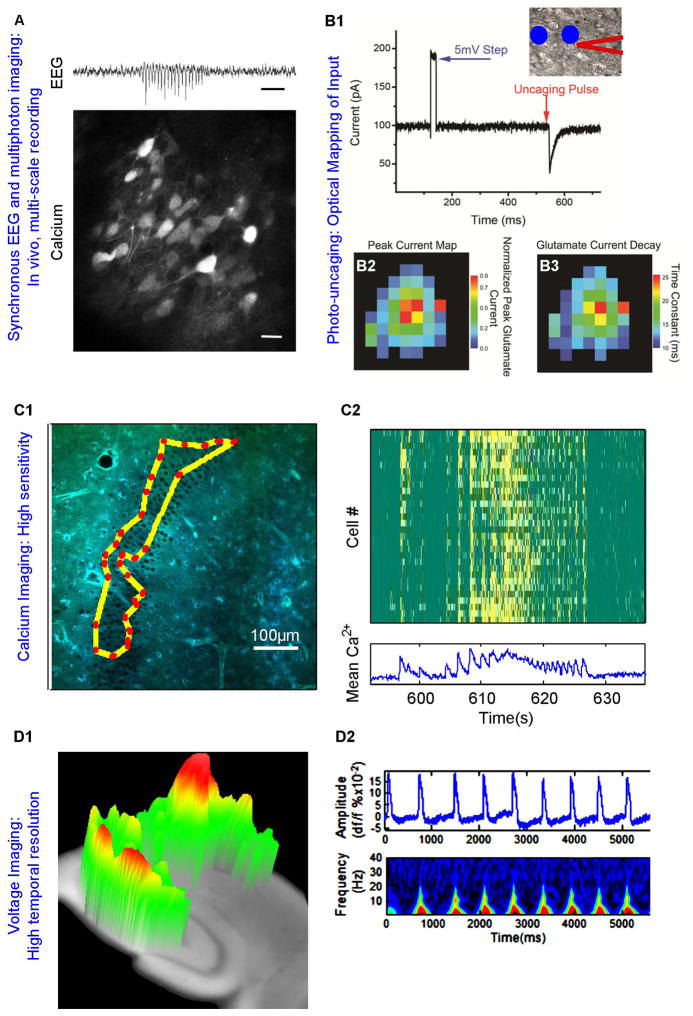
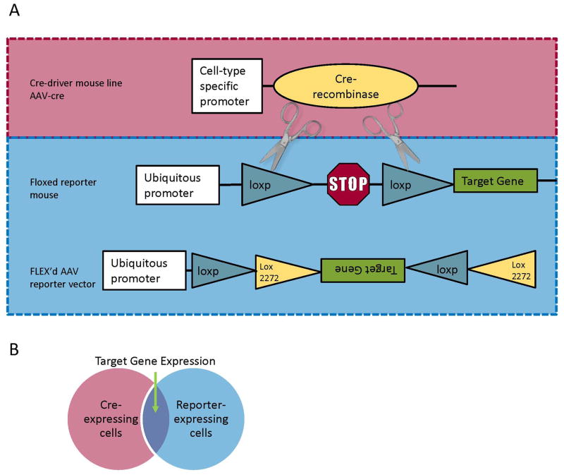
Great advancements have been made in understanding the basic mechanisms of ictogenesis using single-cell electrophysiology (e.g., patch clamp, sharp electrode), large-scale electrophysiology (e.g., electroencephalography [EEG], field potential recording), and large-scale imaging (magnetic resonance imaging [MRI], positron emission tomography [PET], calcium imaging of acetoxymethyl ester [AM] dye-loaded tissue). Until recently, it has been challenging to study experimentally how population rhythms emerge from cellular activity. Newly developed optical imaging technologies hold promise for bridging this gap by making it possible to simultaneously record the many cellular elements that comprise a neural circuit. Furthermore, easily accessible genetic technologies for targeting expression of fluorescent protein-based indicators make it possible to study, in animal models of epilepsy, epileptogenic changes to neural circuits over long periods. In this review, we summarize some of the latest imaging tools (fluorescent probes, gene delivery methods, and microscopy techniques) that can lead to the advancement of cell- and circuit-level understanding of epilepsy, which in turn may inform and improve development of next generation antiepileptic and antiepileptogenic drugs.

Keywords: Imaging; Microscopy; Probes; WONOEP.


Conflict of interest statement

Disclosure

None of the authors has any conflict of interest to disclose.

Figures

Figure 1
Figure 1. Molecular, cellular, and network imaging at the circuit level
A) While simultaneously recording intracranial EEG (top, scale = 1 second), calcium activity can be imaged from neurons transfected with GCaMP6 as seen here in layer 2/3 of visual cortex of stargazer, a mouse model of absence epilepsy (bottom, scale = 20 μm; courtesy of Jochen Meyer, Atul Maheshwari, Stelios Smirnakis and Jeffrey Noebels, Baylor College of Medicine). B1) Differential interference contrast of a cortical astrocyte (center) shown with patch-clamp electrode (outlined in red). Photostimulation sites are indicated by blue circles. Whole-cell recording of an astrocyte voltage-clamped at −80 mV reveals robust glutamate transporter current evoked by photostimulation proximal to the soma. This technique can be repeated to generate maps of peak current (B2) and transporter decay time (B3) for an individual astrocyte. C1) Two-photon Targeted Path Scan (TPS) imaging traces the laser along a path that bisects a predefined subset of cells to rapidly sample many cells across a large area. C2) In this example, TPS was used to sample 30 cells at >30Hz during seizure-like activity in an organotypic slice culture (Lillis and Staley, unpublished). D1) High speed (1.2KHz) voltage-sensitive dye imaging of 4-aminopyridine-evoked epileptiform bursts in enables low resolution 3D visualization of travelling waves hippocampal slices. D2) VSD imaging data can be processed to look at spatial, temporal, and spectral characteristics. Adapted from .
Figure 2
Figure 2. Cre-mediated expression
A) A typical scheme for targeted expression of a genetically encoded fluorescent probe involves cre recombinase-mediated deletion of a loxp-flanked stop codon to achieve expression of the target gene. Cre can be delivered in the form of a transgenic “cre-driver” animal that expresses cre in a subset of cells or an AAV virus. Similarly the “reporter” construct can be a transgenic “floxed” mouse or a FLEX’d virus, both of which constitutively express the target gene following cre-mediated recombination. The “floxed” construct snips out a stop codon upon cre-mediated recombination. The FLEX construct first flips the transgene to the forward direction, and then excises one each of the lox sites to prevent further recombination. B) Target gene expression is achieved only in cells co-expressing cre-recombinase and reporter construct. Typically expression is directed to a set of cells genetically defined by cre expression. When viral constructs are used, spatial organization of expression can also be determined by the site of injection.

References

    1. Claes L, Del-Favero J, Ceulemans B, et al. De Novo Mutations in the Sodium-Channel Gene SCN1A Cause Severe Myoclonic Epilepsy of Infancy. Am J Hum Genet. 2001;68:1327–32. - PMC - PubMed
    1. O’Leary T, Williams AH, Caplan JS, et al. Correlations in ion channel expression emerge from homeostatic tuning rules. Proc Natl Acad Sci. 2013;110:E2645–E2654. - PMC - PubMed
    1. Hüttmann K, Sadgrove M, Wallraff A, et al. Seizures preferentially stimulate proliferation of radial glia-like astrocytes in the adult dentate gyrus: functional and immunocytochemical analysis. Eur J Neurosci. 2003;18:2769–78. - PubMed
    1. Avignone E, Ulmann L, Levavasseur F, et al. Status Epilepticus Induces a Particular Microglial Activation State Characterized by Enhanced Purinergic Signaling. J Neurosci. 2008;28:9133–44. - PMC - PubMed
    1. Danzer SC, He X, Loepke AW, et al. Structural plasticity of dentate granule cell mossy fibers during the development of limbic epilepsy. Hippocampus. 2010;20:113–24. - PMC - PubMed

Publication types

Substances