. 2015 Mar 10:3:e820.
doi: 10.7717/peerj.820. eCollection 2015.

Cell kinetics during regeneration in the sponge Halisarca caerulea: how local is the response to tissue damage?

Affiliations

Cell kinetics during regeneration in the sponge Halisarca caerulea: how local is the response to tissue damage?

Brittany E Alexander et al. PeerJ. .

Abstract

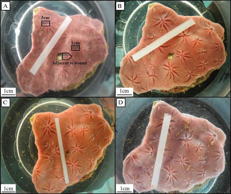
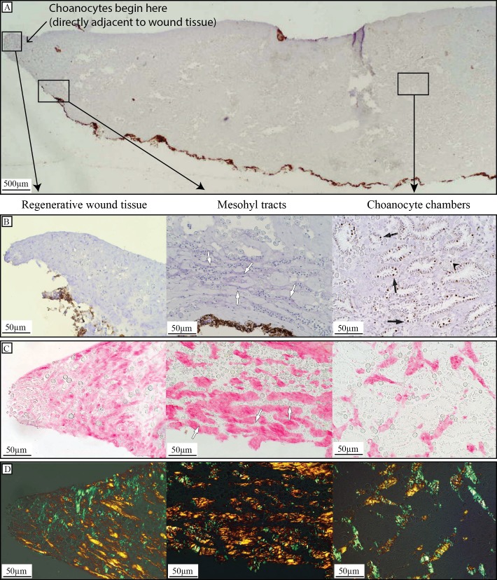
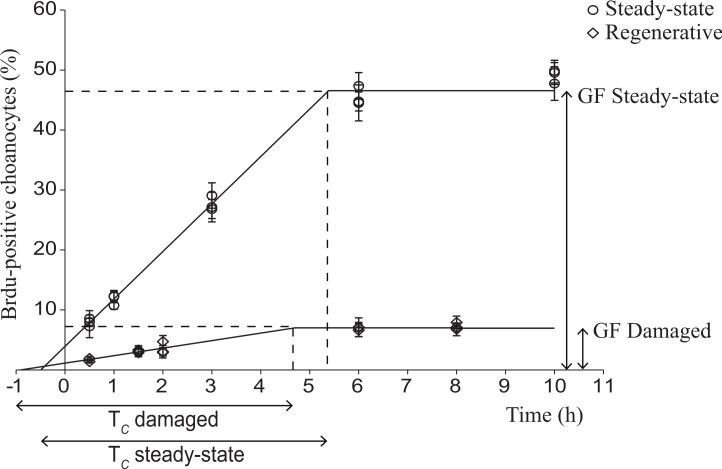
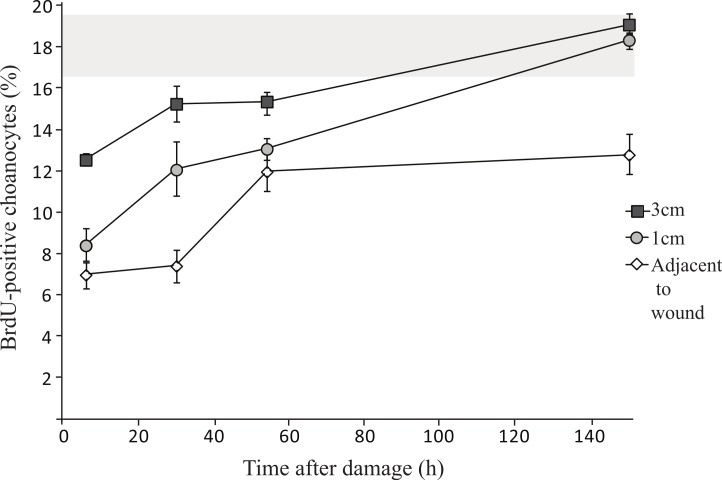
Sponges have a remarkable capacity to rapidly regenerate in response to wound infliction. In addition, sponges rapidly renew their filter systems (choanocytes) to maintain a healthy population of cells. This study describes the cell kinetics of choanocytes in the encrusting reef sponge Halisarca caerulea during early regeneration (0-8 h) following experimental wound infliction. Subsequently, we investigated the spatial relationship between regeneration and cell proliferation over a six-day period directly adjacent to the wound, 1 cm, and 3 cm from the wound. Cell proliferation was determined by the incorporation of 5-bromo-2'-deoxyuridine (BrdU). We demonstrate that during early regeneration, the growth fraction of the choanocytes (i.e., the percentage of proliferative cells) adjacent to the wound is reduced (7.0 ± 2.5%) compared to steady-state, undamaged tissue (46.6 ± 2.6%), while the length of the cell cycle remained short (5.6 ± 3.4 h). The percentage of proliferative choanocytes increased over time in all areas and after six days of regeneration choanocyte proliferation rates were comparable to steady-state tissue. Tissue areas farther from the wound had higher rates of choanocyte proliferation than areas closer to the wound, indicating that more resources are demanded from tissue in the immediate vicinity of the wound. There was no difference in the number of proliferative mesohyl cells in regenerative sponges compared to steady-state sponges. Our data suggest that the production of collagen-rich wound tissue is a key process in tissue regeneration for H. caerulea, and helps to rapidly occupy the bare substratum exposed by the wound. Regeneration and choanocyte renewal are competing and negatively correlated life-history traits, both essential to the survival of sponges. The efficient allocation of limited resources to these life-history traits has enabled the ecological success and diversification of sponges.

Keywords: Cell kinetics; Choanocyte turnover; Collagen; Immunohistochemistry; Modular integration; Regeneration; Sponges; Trade-off.


Conflict of interest statement

Brittany E. Alexander, Ronald Osinga and Jasper M. de Goeij are employees of Porifarma B.V., Poelbos, The Netherlands.

Figures

Figure 1
Figure 1. Regeneration in H. caerulea (A) directly, (B) one day, (C) two days, and (D) six days after wound infliction.
After six days, sponges had completely filled in the bare substrate exposed by the initial wound with a thin layer of regenerative wound tissue. Tissue samples were taken directly adjacent to the wound, 1 cm from the wound, and 3 cm from the wound (A). Tissue samples taken adjacent to the wound were marked with an arrow shape so that the orientation of the tissue could be recognized and histological sections could be made that included the wound area (A). Within each tissue sample, three histological sections were analyzed, each 100 µm apart, represented by the solid black lines (A). Photographs by Brittany Alexander.
Figure 2
Figure 2. Regenerative tissue of H. caerulea.
(A) Cross-section through BrdU- and hematoxylin-stained sponge tissue two days after wound infliction, showing regenerative tissue at the site of the wound, the location of mesohyl tracts containing cells, which were occasionally observed close to the wound, and an area away from the wound containing choanocyte chambers. Choanocyte chambers appeared 250 ± 8.9 µm from the edge of the wound tissue. (B) BrdU immunohistochemistry of a regenerative sponge labeled with BrdU for 6 h. Brdu-positive cells (brown-stained nuclei) were absent from regenerative wound tissue, and cells located in mesohyl tracts were BrdU-negative (blue-stained nuclei, white arrows). Tissue areas away from the wound that had retained their structural integrity contained BrdU-positive choanocytes (black arrows) and occasionally BrdU-positive mesohyl cells (black arrow heads). (C) Picrosirius red staining showed a higher density of collagen in regenerative wound tissue compared to areas farther from the wound. High densities of collagen could also be seen surrounding tracts in the mesohyl containing cells (white arrows). (D) Visualization of picrosirius red staining under cross polarized light revealed thin (green) and thick (orange) collagen fibers in all tissue areas.
Figure 3
Figure 3. Choanocyte cell kinetics in early regenerative and steady-state tissue of H. caerulea.
Steady-state data were obtained from De Goeij and colleagues (2009). In both tissues, the percentage of BrdU-positive choanocytes (mean ± SE) increased over time until a maximum was reached, representing the growth fraction (GF), i.e., the percentage of choanocytes involved in proliferation. The growth fraction of choanocytes in early regenerative tissue was substantially lower than the growth fraction of choanocytes in steady-state tissue. The duration of the linear increase represents the length of the cell cycle, which was similar in regenerative and steady-state sponges. The lines are the least squares fit obtained using the conditions of the ‘one population model’ described by Nowakowski and colleagues (1989).
Figure 4
Figure 4. Changes in choanocyte proliferation rates over time in H. caerulea during regeneration.
There are significantly less proliferative choanocytes closer to the wound compared to 1 cm and 3 cm from the wound (mean ± SE). The percentage of proliferative choanocytes increases over time in each tissue area. The percentages of proliferative choanocytes six days after damage, in tissue located 1 cm, and 3 cm from the wound, are comparable to the percentage of proliferative choanocytes found in steady-state H. caerulea specimens (grey area represents steady-state mean ± SE; data taken from Alexander and colleagues (2014)).

References

    1. Aldewachi H, Wright N, Appleton D, Watson A. Effect of starvation and refeeding on cell-population kinetics in rat small bowel mucosa. Journal of Anatomy. 1975;119:105–121. - PMC - PubMed
    1. Alexander BE, Liebrand K, Osinga R, Van der Geest HG, Admiraal W, Cleutjens JPM, Schutte B, Verheyen F, Ribes M, Van Loon E, de Goeij JM. Cell turnover and detritus production in marine sponges from tropical and temperate benthic ecosystems. PLoS ONE. 2014;9(10):e820. doi: 10.1371/journal.pone.0109486. - DOI - PMC - PubMed
    1. Antcliffe JB, Callow RHT, Brasier MD. Giving the early fossil record of sponges a squeeze. Biological Reviews. 2014;89:972–1004. doi: 10.1111/brv.12090. - DOI - PubMed
    1. Ayling AL. Growth and regeneration rates in thinly encrusting demospongiae from temperate waters. Biological Bulletin. 1983;165:343–352. doi: 10.2307/1541200. - DOI - PubMed
    1. Bell JJ. Regeneration rates of a sublittoral demosponge. Journal of the Marine Biology Association UK. 2002;82:169–170. doi: 10.1017/S0025315402005465. - DOI

LinkOut - more resources