. 2015 Mar 10:3:e831.
doi: 10.7717/peerj.831. eCollection 2015.

Subject-specific body segment parameter estimation using 3D photogrammetry with multiple cameras

Affiliations

Subject-specific body segment parameter estimation using 3D photogrammetry with multiple cameras

Kathrin E Peyer et al. PeerJ. .

Abstract

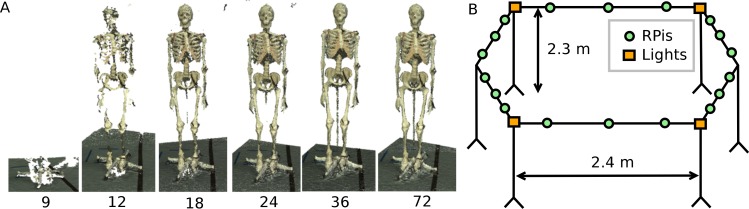
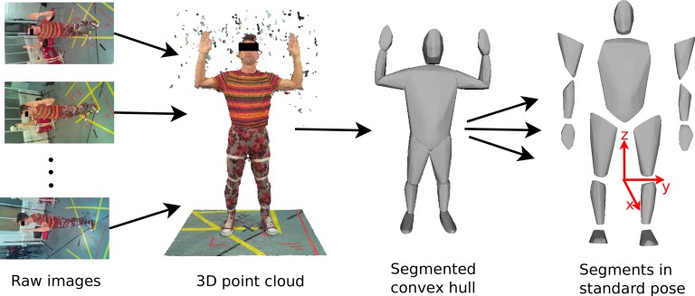
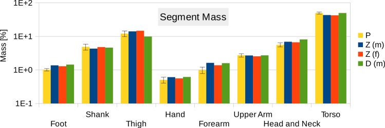
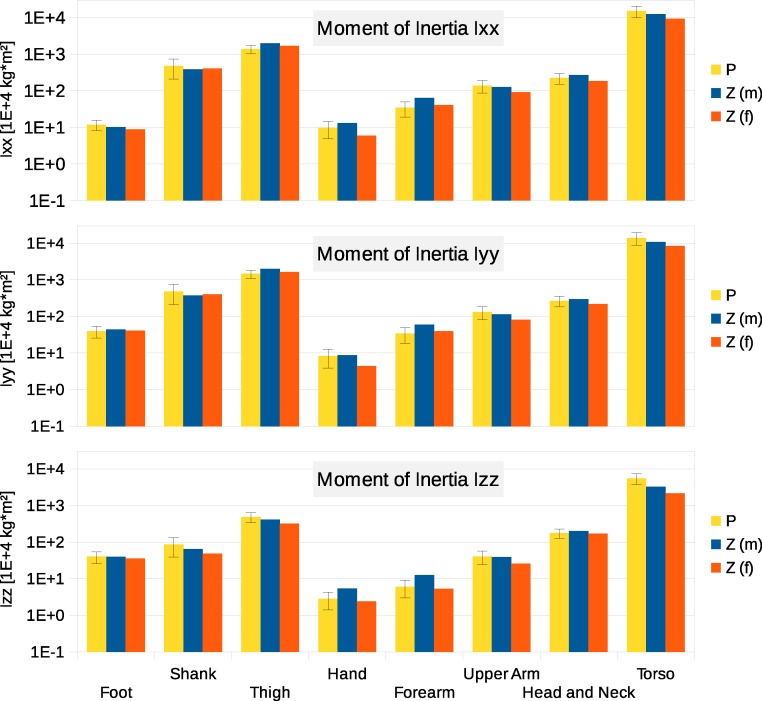
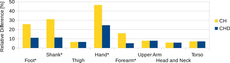
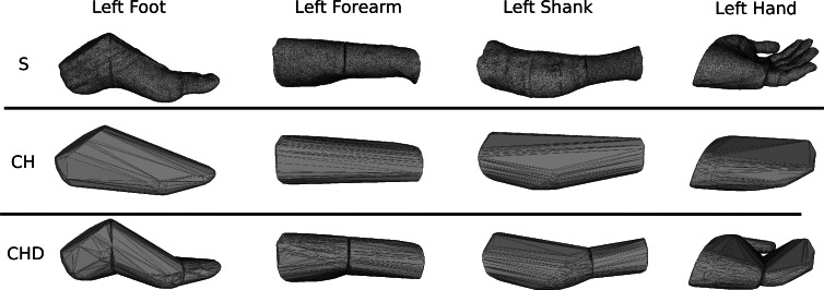
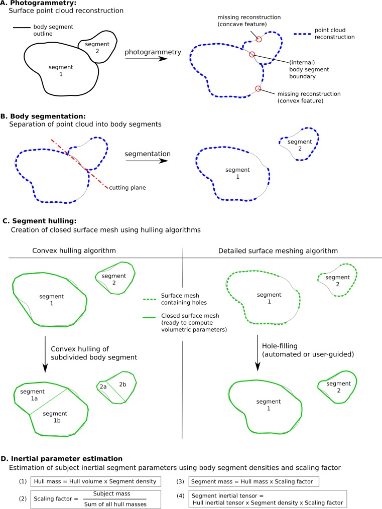
Inertial properties of body segments, such as mass, centre of mass or moments of inertia, are important parameters when studying movements of the human body. However, these quantities are not directly measurable. Current approaches include using regression models which have limited accuracy: geometric models with lengthy measuring procedures or acquiring and post-processing MRI scans of participants. We propose a geometric methodology based on 3D photogrammetry using multiple cameras to provide subject-specific body segment parameters while minimizing the interaction time with the participants. A low-cost body scanner was built using multiple cameras and 3D point cloud data generated using structure from motion photogrammetric reconstruction algorithms. The point cloud was manually separated into body segments, and convex hulling applied to each segment to produce the required geometric outlines. The accuracy of the method can be adjusted by choosing the number of subdivisions of the body segments. The body segment parameters of six participants (four male and two female) are presented using the proposed method. The multi-camera photogrammetric approach is expected to be particularly suited for studies including populations for which regression models are not available in literature and where other geometric techniques or MRI scanning are not applicable due to time or ethical constraints.

Keywords: Biomechanics; Body segment parameters; Geometric modelling; Photogrammetry; Structure from motion; Subject-specific estimation.


Conflict of interest statement

The authors declare there are no competing interests.

Figures

Figure 1
Figure 1. Body scanner design.
(A) Point cloud reconstruction with varying number of cameras. (B) Schematic representation of the RPi scanner design.
Figure 2
Figure 2. Image processing work flow.
Images from the RPI scanner are converted to 3D point clouds which are then scaled and segmented manually. Subsequently, convex hulling is used to produce a surface mesh around each body segment.
Figure 3
Figure 3. Segment mass (as % of body mass).
P, Average value of all six participants (error bars show standard deviation). Foot mass adjusted by a factor of 0.51 to compensate for volume overestimation due to wearing shoes. Z(m), Male average values reported by Zatsiorsky; Z(f), Female average values reported by Zatsiorsky (Leva, 1996; Zatsiorsky, 2002); D(m), Male average values by Dempster (via Zatsiorsky) (Dempster, 1955; Zatsiorsky, 2002).
Figure 4
Figure 4. Moment of inertia in (104 kg m2)
P, Average value of all six participants (error bars show standard deviation). Foot moment of inertia adjusted by a factor of 0.51 to compensate for volume overestimation due to wearing shoes. Z(m), Male average values reported by Zatsiorsky; Z(f), Female average values reported by Zatsiorsky (Leva, 1996; Zatsiorsky, 2002). The definition of the coordinate system is shown in Fig. 2.
Figure 5
Figure 5. Absolute values of products of inertia in (104 kg m2).
The absolute values of Ixy, Ixz and Iyz are shown together with a positive error bar (negative error bar is symmetrical) equal to one standard deviation. The signed values are reported in Supplemental Information (Tables S2–S4). The Ixy value of the hand is smaller than 103 kg m2 and is not displayed. Foot products of inertia adjusted by a factor of 0.51 to compensate for volume overestimation due to wearing shoes.
Figure 6
Figure 6. Centre of mass along the longitudinal axis.
P, Average value of all six participants (error bars show standard deviation). Z(m, male; f, female): Average values by Zatsiorsky, adjusted by de Leva. The CoM is given as % of the segment length. The definition of the segments and reference points are given in Supplemental Information Table S1 - Exceptions: * Foot of participants: Heel and toe end point of participant’s shoes instead of foot. ** Forearm and Upper Arm of Z: Elbow reference point is the elbow joint centre instead of the Olecranon (Leva, 1996; Zatsiorsky, 2002).
Figure 7
Figure 7. CoM shift from the anatomical longitudinal axis in the transverse (xy) plane.
Average values of all six participants are shown (error bars show standard deviation). Due to mirror-symmetry, the y-values of the segments on the left- and right-hand side have opposite signs. To calculate the average, the sign of the segments on the left-hand side was inverted. The CoM is given as % of the segment length. The data of the foot is not included due to the participants wearing shoes.
Figure 8
Figure 8. Visible human surface mesh.
(A) High-resolution surface mesh. (B) Convex hull mesh.
Figure 9
Figure 9. Segment volume overestimation of the hulled mesh versus the high-resolution surface mesh of the visible human.
Data shown as the relative difference of the hull with respect to the original mesh. CH, Convex hull of body segment; CHD, Convex hull of divided body segments (only segments indicated with an * were subdivided, see Fig. 10).
Figure 10
Figure 10. Subdivision of the body segments with large curvature.
The first row (S) shows the high-resolution surface mesh, the second row (CH) the convex hull of the whole body segment, and the bottom row (CHD) the convex hulls of the subdivided body segments.
Figure 11
Figure 11. Male visible human segment mass (as % of body mass) of the high-resolution mesh, convex hull, regression model and average values.
S, High-resolution surface mesh; CH, Convex Hull of whole body segments; CHD, Convex Hull with subdivided body segments (only segments indicated with an * were subdivided as shown in Fig. 10); ZR, Values predicted using Zatsiosrky’s linear regression model (using weight and height); Z, Male average values reported by Zatsiorsky; D, Male average values reported by Dempster (Dempster, 1955; Leva, 1996; Zatsiorsky, 2002).
Figure 12
Figure 12. Methodology to estimate subject-specific body segment parameters using photogrammetry.
(A) Photogrammetry; (B) Body segmentation; (C) Segment hulling; (D) Inertial parameter estimation.

References

    1. Agarwal S, Snavely N, Simon I, Seitz SM, Szeliski R. Building Rome in a day. IEEE international conference on computer vision; Piscataway. 2009. pp. 72–79.
    1. Bates KT, Manning PL, Margetts L, Sellers WI. Sensitivity analysis in evolutionary robotic simulations of bipedal dinosaur running. Journal of Vertebrate Paleontology. 2010;30:458–466. doi: 10.1080/02724630903409329. - DOI
    1. Bauer JJ, Pavol MJ, Snow CM, Hayes WC. MRI-derived body segment parameters of children differ from age-based estimates derived using photogrammetry. Journal of Biomechanics. 2007;40:2904–2910. doi: 10.1016/j.jbiomech.2007.03.006. - DOI - PubMed
    1. Bernardini F, Mittleman J, Rushmeier H, Silva C, Taubin G. The ball-pivoting algorithm for surface reconstruction. IEEE Transactions on Visualization and Computer Graphics. 1999;5:349–359. doi: 10.1109/2945.817351. - DOI
    1. Bonnechère B, Jansen B, Salvia P, Bouzahouene H, Sholukha V, Cornelis J, Rooze M, Van Sint Jan S. Determination of the precision and accuracy of morphological measurements using the KinectTM sensor: comparison with standard stereophotogrammetry. Ergonomics. 2014;57:622–631. doi: 10.1080/00140139.2014.884246. - DOI - PubMed

LinkOut - more resources