. 2015 Mar 17;10(3):e0118550.
doi: 10.1371/journal.pone.0118550. eCollection 2015.

Analysis of neurogenesis during experimental autoimmune encephalomyelitis reveals pitfalls of bioluminescence imaging

Affiliations

Analysis of neurogenesis during experimental autoimmune encephalomyelitis reveals pitfalls of bioluminescence imaging

Ilya Ayzenberg et al. PLoS One. .

Abstract

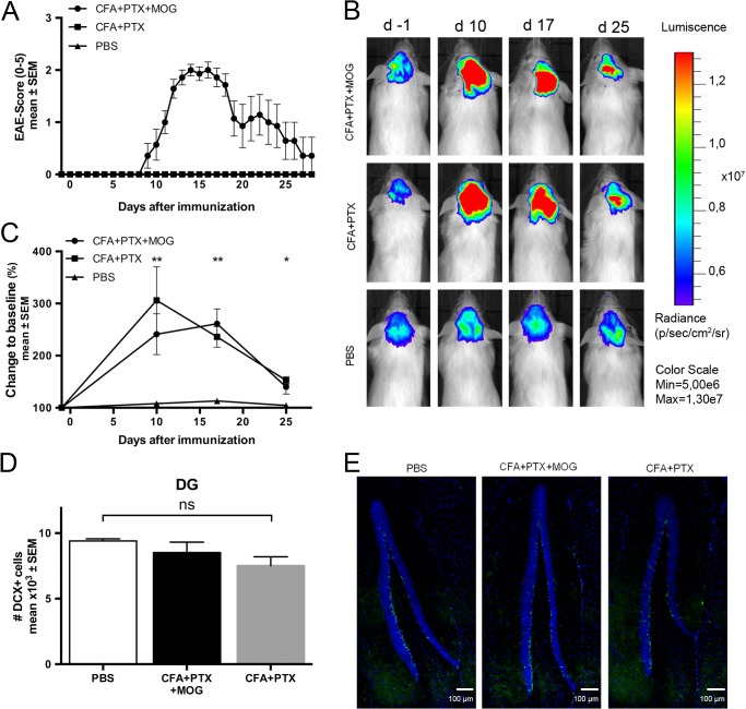
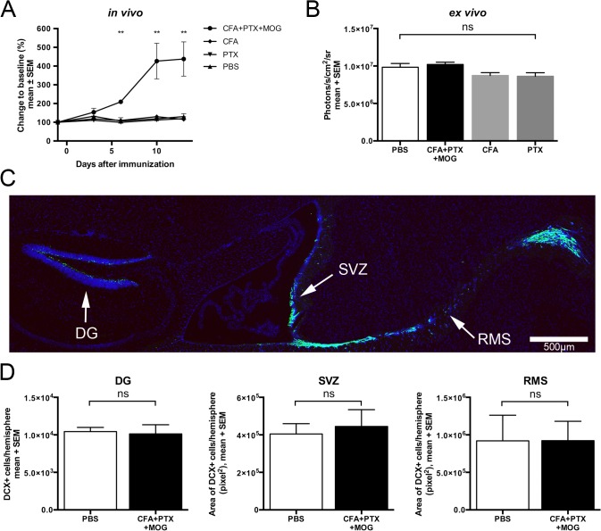
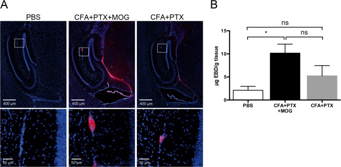
Bioluminescence imaging is a sensitive approach for longitudinal neuroimaging. Transgenic mice expressing luciferase under the promoter of doublecortin (DCX-luc), a specific marker of neuronal progenitor cells (NPC), allow monitoring of neurogenesis in living mice. Since the extent and time course of neurogenesis during autoimmune brain inflammation are controversial, we investigated neurogenesis in MOG-peptide induced experimental allergic encephalomyelitis (EAE) using DCX-luc reporter mice. We observed a marked, 2- to 4-fold increase of the bioluminescence signal intensity 10 days after EAE induction and a gradual decline 1-2 weeks thereafter. In contrast, immunostaining for DCX revealed no differences between EAE and control mice 2 and 4 weeks after immunization in zones of adult murine neurogenesis such as the dentate gyrus. Ex vivo bioluminescence imaging showed similar luciferase expression in brain homogenates of EAE and control animals. Apart from complete immunization including MOG-peptide also incomplete immunization with complete Freund´s adjuvant and pertussis toxin resulted in a rapid increase of the in vivo bioluminescence signal. Blood-brain barrier (BBB) leakage was demonstrated 10 days after both complete and incomplete immunization and might explain the increased bioluminescence signal in vivo. We conclude, that acute autoimmune inflammation in EAE does not alter neurogenesis, at least at the stage of DCX-expressing NPC. Effects of immunization on the BBB integrity must be considered when luciferase is used as a reporter within the CNS during the active stage of EAE. Models with stable CNS-restricted luciferase expression could serve as technically convenient way to evaluate BBB integrity in a longitudinal manner.


Conflict of interest statement

Competing Interests: IK has received honoraria for consultancy/speaking and travel reimbursement from Bayer Health Care, Biogen Idec, Chugai, and Novartis Pharmaceuticals and has received research grants from Bayer Health Care, Biogen Idec, and Novartis Pharmaceuticals. The other authors declare that they have no competing interests. This does not alter the authors' adherence to PLOS ONE policies on sharing data and materials.

Figures

Fig 1
Fig 1. In vivo bioluminescence imaging of neurogenesis in mice with EAE and after incomplete immunization.
(A) Experimental autoimmune encephalomyelitis (EAE) was induced in doublecortin (DCX)-luc reporter mice by immunization with 200 μg MOG35–55 peptide emulsified in CFA+PTX (n = 7). Control mice received incomplete immunization (CFA PTX; n = 5) or PBS (n = 6). The clinical course of EAE was followed for 28 days. (B and C) Bioluminescence of the brain was recorded from minute 5 to 20 after i.p. injection of 150 mg/kg D-Luciferin. The maximum photon flux integrated over 59 seconds is shown. (B) illustrates representative examples, (C) temporal changes of the average bioluminescence signal at 4 time points. Mean + SEM is shown. *p<0.05, **p<0.01 (ANOVA). (D and E) Neurogenesis in the dentate gyrus (DG) 28 days after immunization. DCX positive cells in the DG were counted and presented as the total number of positive cells per hemisphere (mean ± SEM; statistical analysis by ANOVA).
Fig 2
Fig 2. DCX-expression and ex vivo bioluminescence remain constant despite increased in vivo bioluminescence after EAE induction.
(A) In vivo bioluminescence imaging of the brain recorded from minute 5 to 20 after i.p. injection of 150 mg/kg D-Luciferin in DCX-luc mice with antigen-specific (200 μg MOG35–55+CFA+PTX, n = 6) or incomplete (CFA only, n = 4; PTX only, n = 4) immunization and in the PBS control group (n = 5). The maximum photon flux integrated over 59 seconds is shown. Only mice with antigen-specific immunization developed clinical signs of EAE (not shown). Mice were sacrificed after 14 days and brains were used for ex vivo detection of luciferase activity. (B) Brain hemispheres were homogenized and luciferase activity measured by addition of excess D-Luciferin and ATP. (C and D) DCX-luc mice were immunized as detailed above (PBS, n = 6; CFA+PTX+MOG, n = 6) and perfused after 14 days at the peak of EAE. Sagittal brain sections were immunostained for DCX (shown in green) and analyzed in the dentate gyrus (DG), subventricular zone (SVZ), and rostral migratory stream (RMS). (C) A representative example is shown (PBS-treated). (D) DCX positive cells in the DG were counted and presented as the total number of positive cells per hemisphere. DCX positive cells in the SVZ and the RMS are shown as the total area (pixel2) of positive cells per hemisphere. Results are presented as mean ± SEM per group. **p<0.01 (ANOVA in A, B; Mann Whitney U test in D).
Fig 3
Fig 3. Disruption of the blood-brain-barrier after antigen-specific and incomplete immunization.
(A) At day 10 after antigen-specific and incomplete immunization (CFA+PTX+MOG, n = 3; CFA+PTX, n = 4; PBS n = 4) mice were intravenously injected with 4% Evans blue dye (EBD) and sacrificed after 20 minutes of EBD circulation. Fluorescence microscopy revealed an increased blood-brain barrier (BBB) permeability in both immunized groups, most prominent between the DG and the thalamus, in the choroid plexus and around small vessels (insets). (B) Measurement of extracted EBD from brain parenchyma, expressed as μg per g tissue confirms leakage of the BBB after immunization. Results are presented as a mean ± SEM per group. *p<0.05 (ANOVA).

References

    1. Hochgrafe K, Mandelkow EM. Making the brain glow: in vivo bioluminescence imaging to study neurodegeneration. Mol Neurobiol. 2013;47(3):868–82. Epub 2012/11/30. 10.1007/s12035-012-8379-1 PubMed PMID: . - DOI - PubMed
    1. Luo J, Ho PP, Buckwalter MS, Hsu T, Lee LY, Zhang H, et al. Glia-dependent TGF-beta signaling, acting independently of the TH17 pathway, is critical for initiation of murine autoimmune encephalomyelitis. J Clin Invest. 2007;117(11):3306–15. Epub 2007/10/30. 10.1172/JCI31763 PubMed PMID: . - DOI - PMC - PubMed
    1. Luo J, Ho P, Steinman L, Wyss-Coray T. Bioluminescence in vivo imaging of autoimmune encephalomyelitis predicts disease. J Neuroinflammation. 2008;5:6 Epub 2008/02/02. 1742-2094-5-6 [pii] 10.1186/1742-2094-5-6 PubMed PMID: . - DOI - PMC - PubMed
    1. Kim H, Walczak P, Kerr C, Galpoththawela C, Gilad AA, Muja N, et al. Immunomodulation by transplanted human embryonic stem cell-derived oligodendroglial progenitors in experimental autoimmune encephalomyelitis. Stem Cells. 2012;30(12):2820–9. Epub 2012/09/06. 10.1002/stem.1218 PubMed PMID: - DOI - PMC - PubMed
    1. Sher F, Amor S, Gerritsen W, Baker D, Jackson SL, Boddeke E, et al. Intraventricularly injected Olig2-NSCs attenuate established relapsing-remitting EAE in mice. Cell Transplant. 2012;21(9):1883–97. Epub 2012/04/04. 10.3727/096368911X637443 PubMed PMID: . - DOI - PubMed

Publication types

LinkOut - more resources