. 2015 May 15;593(10):2295-310.
doi: 10.1113/JP270045. Epub 2015 Apr 13.

Inhibition of presynaptic calcium transients in cortical inputs to the dorsolateral striatum by metabotropic GABA(B) and mGlu2/3 receptors

Affiliations

Inhibition of presynaptic calcium transients in cortical inputs to the dorsolateral striatum by metabotropic GABA(B) and mGlu2/3 receptors

David A Kupferschmidt et al. J Physiol. .

Abstract

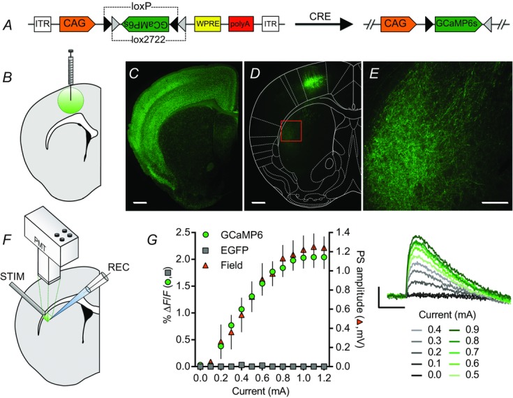
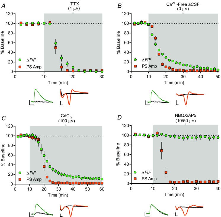
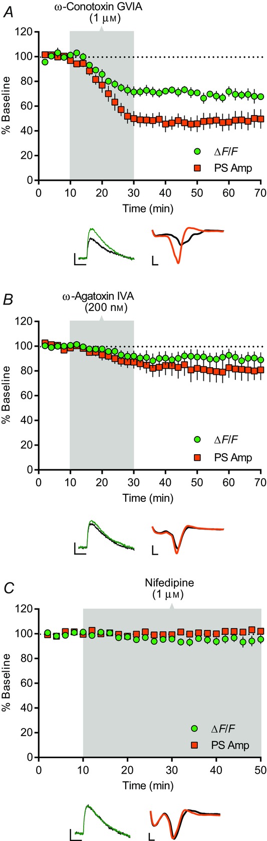
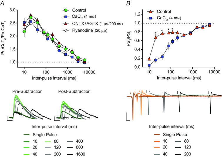
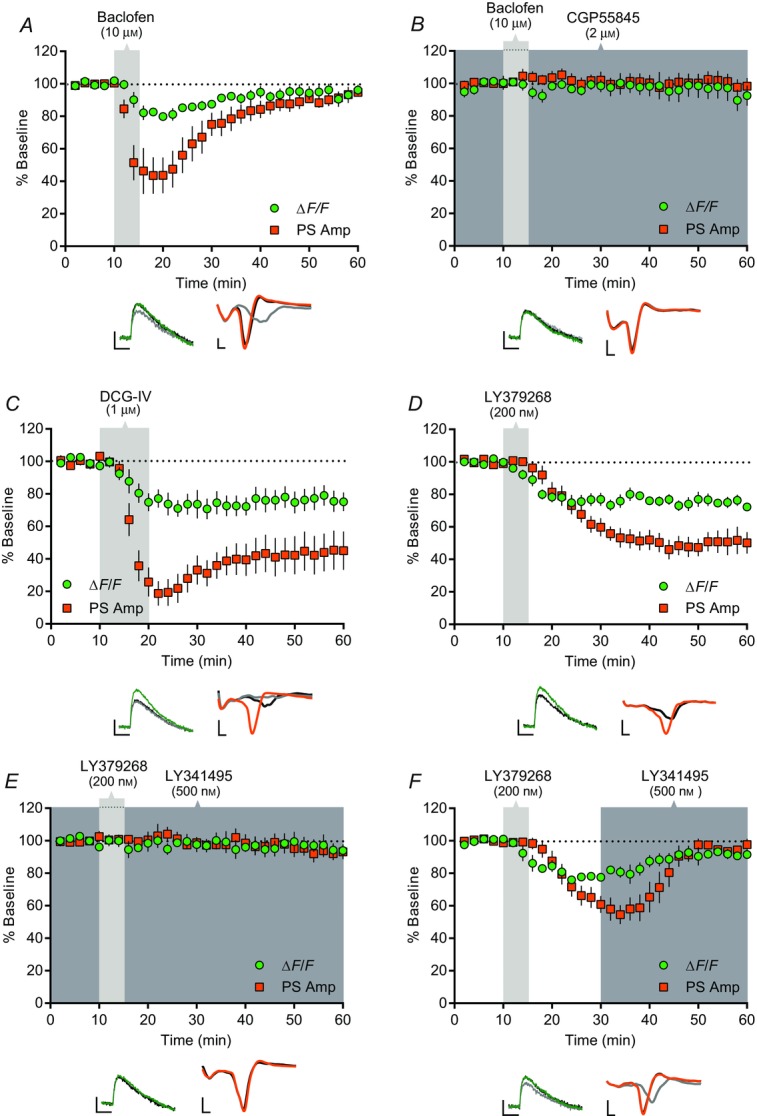
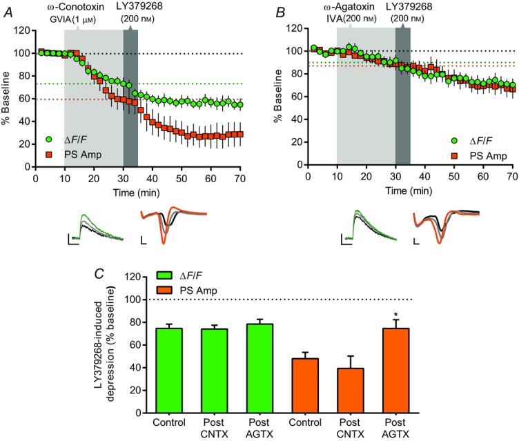
Cortical inputs to the dorsolateral striatum (DLS) are dynamically regulated during skill learning and habit formation, and are dysregulated in disorders characterized by impaired action control. Therefore, a mechanistic investigation of the processes regulating corticostriatal transmission is key to understanding DLS-associated circuit function, behaviour and pathology. Presynaptic GABA(B) and group II metabotropic glutamate (mGlu2/3) receptors exert marked inhibitory control over corticostriatal glutamate release in the DLS, yet the signalling pathways through which they do so are unclear. We developed a novel approach using the genetically encoded calcium (Ca(2+) ) indicator GCaMP6 to assess presynaptic Ca(2+) in corticostriatal projections to the DLS. Using simultaneous photometric presynaptic Ca(2+) and striatal field potential recordings, we report that relative to P/Q-type Ca(2+) channels, N-type channels preferentially contributed to evoked presynaptic Ca(2+) influx in motor cortex projections to, and excitatory transmission in, the DLS. Activation of GABA(B) or mGlu2/3 receptors inhibited both evoked presynaptic Ca(2+) transients and striatal field potentials. mGlu2/3 receptor-mediated depression did not require functional N-type Ca(2+) channels, but was attenuated by blockade of P/Q-type channels. These findings reveal presynaptic mechanisms of inhibitory modulation of corticostriatal function that probably contribute to the selection and shaping of behavioural repertoires.


Figures

Figure 1
Figure 1
GCaMP6s expression in corticostriatal inputs to dorsolateral striatum A, schematic diagram of the AAV-FLEX-GCaMP6s construct. In the presence of Cre recombinase, permanent recombination of two pairs of loxP and lox2272 sites changes the orientation of the GCaMP6 sequence from anti-sense to sense with respect to its promoter, CAG. B, schematic diagram of an AAV-FLEX-GCaMP6s injection into M1. C, cre-mediated expression of EGFP preferentially in cortical pyramidal neurons (and cortical glia; Gorski et al. 2002) in a striatal-containing coronal brain slice from an Emx1Cre;CAG-floxedSTOP-ZsGreen mouse. D and E, cre-mediated GCaMP6s expression in M1 cortical pyramidal cells (D) and presynaptic corticostriatal elements in the DLS (D, E) of an Emx1Cre mouse injected with AAV-FLEX-GCaMP6s into M1. The area shown in E is indicated by the box in D. F, schematic diagram of brain slice photometry and electrophysiology recordings. A concentric bipolar stimulating electrode placed at the border of the DLS and overlying white matter evoked striatal fluorescent transients detected by a photomultiplier tube (PMT) and striatal field potentials detected by a glass recording electrode. G, current–response curves of 100 μs electrical pulse-evoked fluorescence and field potentials recorded in the DLS of Emx1Cre mice injected with AAV-FLEX-GCaMP6s or AAV-FLEX-EGFP. GCaMP6s- (green circles, n = 6) but not EGFP-injected mice (grey squares, n = 6) showed evoked fluorescent transients that scaled with stimulation current amplitude and population spike (PS) amplitude (orange triangles, n = 6) (traces on right; average of four evoked responses per current amplitude). Scale bars of fluorescence images: 500 μm (C, D), 200 μm (E). Scale bars of evoked PreCaTs from AAV-FLEX-GCaMP6s-injected Emx1Cre mice: 1% ΔF/F, 0.5 s (G).
Figure 2
Figure 2
Characterization of corticostriatal PreCaTs in dorsolateral striatum Time course of the effects of TTX, Ca2+-free aCSF, CdCl2 and NBQX/AP5 on electrical pulse-evoked photometric PreCaT and electrophysiological field potential recordings in the DLS. PreCaT amplitude is expressed as % baseline ΔF/F (green circles) and population spike amplitude is expressed as % baseline PS Amp (orange squares). Shaded areas correspond to periods of drug application. Representative traces from PreCaT (left, average of four evoked responses) and field potential recordings (right, average of eight evoked responses) are shown below each graph. A, bath application of the sodium channel blocker, TTX (1 μm), for 20 min abolished PreCaTs and PSs. B, bath application of Ca2+-free aCSF for 40 min abolished PreCaTs and PSs. C, bath application of the non-selective Ca2+ channel blocker, CdCl2 (100 μm), for 50 min dramatically reduced PreCaT amplitude and abolished PSs. D, bath application of the AMPA and NMDA receptor antagonists, NBQX (10 μm) and AP5 (50 μm), for 30 min had no effect on PreCaT amplitude, but abolished PSs. Trace time points: green/orange, 10 min; black, 70 min (A), 50 min (B), 60 min (C), 40 min (D). Trace scale bars: PreCaTs, 1% ΔF/F, 0.5 s; field potentials, 0.5 mV, 1 ms.
Figure 3
Figure 3
VGCC contribution to corticostriatal PreCaTs and transmission Time course of the effects of ω-conotoxin GVIA, ω-agatoxin IVA and nifedipine on electrical pulse-evoked PreCaT (ΔF/F) and field potential (PS Amp) recordings in the DLS. Shaded areas correspond to periods of drug application. Representative traces from PreCaT (left, average of four evoked responses) and field potential recordings (right, average of eight evoked responses) are shown below each graph. A, bath application of the N-type VGCC blocker, ω-conotoxin GVIA (1 μm), for 20 min suppressed PS amplitude. B, bath application of the P/Q-type VGCC blocker, ω-agatoxin IVA (200 nm), for 20 min modestly suppressed PS amplitude. C, bath application of the L-type VGCC blocker, nifedipine (1 μm), for 40 min had no effect on PreCaT or PS amplitude. Trace time points: green/orange, 10 min; black, 70 min (A, B), 50 min (C). Trace scale bars: PreCaTs, 1% ΔF/F, 0.5 s; field potentials, 0.5 mV, 1 ms.
Figure 4
Figure 4
Paired-pulse ratios of corticostriatal PreCaTs and transmission The ratio of two PreCaTs (PreCaT2/PreCaT1) (A) or two population spikes (PS2/PS1) (B) evoked by two electrical pulses separated by IPIs of 10, 20, 40, 80, 120, 200, 400, 800, 3000 and 6000 ms (IPIs presented on a log scale). A, the effects of CaCl2, ω-conotoxin GVIA (CNTX) with ω-agatoxin IVA (AGTX), and ryanodine on PreCaT PPRs were assessed relative to control conditions. The evoked PreCaT amplitude observed in response to the second of two paired electrical pulses was robustly enhanced relative to the PreCaT amplitude evoked by a single pulse at the majority of IPIs tested (i.e. PPF of PreCaT amplitude, green circles, n = 7). PPF in the presence of high [Ca2+] aCSF (4 mm CaCl2; blue squares, n = 4), following co-application of ω-conotoxin GVIA and ω-agatoxin IVA (1 μm and 200 nm; red triangles, n = 4), or following ryanodine (20 μm; white diamonds, n = 4) was indistinguishable from control conditions (green circles, n = 7). Representative traces of control PreCaTs in response to single- and paired-pulse stimulations, before (left) and following (right) subtraction of the corresponding single-pulse response, are shown below (average of four evoked responses per IPI). Trace scale bars, 1% ΔF/F, 0.5 s. B, the effect of CaCl2 on PS PPRs was assessed relative to control conditions. The evoked PS amplitude observed in response to the second of two paired electrical pulses was depressed relative to the PS amplitude evoked by the first pulse at the majority of IPIs tested (i.e. PPD of PS amplitude, orange triangles, n = 7). PPD in the presence of high [Ca2+] aCSF (4 mm CaCl2; blue squares, n = 5) was enhanced at 20, 40, 80 and 120 ms IPIs relative to control conditions (orange triangles, n = 7). Representative traces of control field recordings in response to single- and paired-pulse stimulations are shown below (average of two evoked responses per IPI). Trace scale bars, 0.5 mV, 10 ms.
Figure 5
Figure 5
GABAB- and mGlu2/3 receptor-mediated depression of corticostriatal PreCaTs and transmission Time course of the effects of baclofen, DCG-IV and LY379268 on electrical stimulation-evoked PreCaT (ΔF/F) and field potential (PS Amp) recordings in the DLS. Shaded areas correspond to periods of drug application. Representative traces from PreCaT (left, average of four evoked responses) and field potential recordings (right, average of eight evoked responses) are shown below each graph. A, bath application of the GABAB receptor agonist, baclofen (10 μm), for 5 min transiently suppressed both PreCaT and PS amplitude. B, pre-application of the GABAB receptor antagonist, CGP55845 (2 μm), prevented baclofen- (10 μm) induced suppression of PreCaT and PS amplitude. C, bath application of the mGlu2/3 receptor agonist, DCG-IV (1 μm), for 10 min suppressed both PreCaT and PS amplitude. D, bath application of the mGlu2/3 receptor agonist, LY379268 (200 nm), for 5 min suppressed both PreCaT and PS amplitude. E, pre-application of the mGlu2/3 receptor antagonist, LY341495 (500 nm), prevented LY379268- (200 nm) induced suppression of PreCaT and PS amplitude. F, bath application of LY341495 (500 nm) for 30 min reversed LY379268- (200 nm) induced suppression of PreCaT and PS amplitude. Trace time points: green/orange, 10 min; grey, 20 min (A), 22 min (C), 30 min (F); black, 60 min. Trace scale bars: PreCaTs, 1% ΔF/F, 0.5 s; field potentials: 0.5 mV, 1 ms.
Figure 6
Figure 6
P/Q-type VGCCs contribute to mGlu2/3 receptor-mediated depression A and B, time course of the effects of ω-conotoxin GVIA (CNTX) or ω-agatoxin IVA (AGTX) on LY379268-induced depression of electrical stimulation-evoked PreCaT (ΔF/F) and field potential (PS Amp) recordings in the DLS. Shaded areas correspond to periods of drug application. The dotted lines denote the average % baseline ΔF/F (green line) and PS Amp (orange line) achieved at termination of VGCC blocker application; these values served as the baselines to which the effects of subsequent LY379268 application were compared. Representative traces from PreCaT (left, average of four evoked responses) and field potential recordings (right, average of eight evoked responses) are shown below each graph. A, pre-application of the N-type VGCC blocker, ω-conotoxin GVIA (1 μm), for 20 min had no effect on LY379268- (200 nm) induced suppression of PreCaT and PS amplitude. B, pre-application of the P/Q-type VGCC blocker, ω-agatoxin IVA (200 nm), for 20 min attenuated LY379268- (200 nm) induced suppression of PS amplitude. LY379268-induced suppression of PreCaT amplitude was unaltered by pre-application of ω-agatoxin IVA. Trace time points: green/orange, 10 min; grey, 30 min; black, 70 min. Trace scale bars: PreCaTs, 1% ΔF/F, 0.5 s; field potentials: 0.5 mV, 1 ms. C, summary of LY379268-induced inhibition of PreCaT and PS amplitude with or without pre-application of VGCC blockers. Inhibition of PS amplitude by LY379268 after P/Q-type VGCC blockade (Post AGTX) was significantly less than that observed after LY379268 alone (Control); *P < 0.05.

Similar articles

Cited by

References

    1. Allen MD. Zhang J. Subcellular dynamics of protein kinase A activity visualized by FRET-based reporters. Biochem Biophys Res Commun. 2006;348:716–721. - PubMed
    1. Akopian G, Musleh W, Smith R. Walsh JP. Functional state of corticostriatal synapses determines their expression of short- and long-term plasticity. Synapse. 2000;38:271–280. - PubMed
    1. Amaral DG. Dent JA. Development of the mossy fibers of the dentate gyrus: I. A light and electron microscopic study of the mossy fibers and their expansions. J Comp Neurol. 1981;195:51–86. - PubMed
    1. Atwood BK, Kupferschmidt DA. Lovinger DM. Opioids induce dissociable forms of long-term depression of excitatory inputs to the dorsal striatum. Nat Neurosci. 2014;17:540–548. - PMC - PubMed
    1. Atwood BK, Lovinger DM. Mathur BN. Presynaptic long-term depression mediated by Gi/o-coupled receptors. Trends Neurosci. 2014;37:663–673. - PMC - PubMed

Publication types

Substances

LinkOut - more resources