Detailed analysis of the human mitochondrial contact site complex indicate a hierarchy of subunits
- PMID: 25781180
- PMCID: PMC4363703
- DOI: 10.1371/journal.pone.0120213
Detailed analysis of the human mitochondrial contact site complex indicate a hierarchy of subunits
Abstract
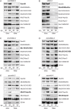
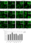
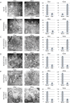
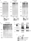
Mitochondrial inner membrane folds into cristae, which significantly increase its surface and are important for mitochondrial function. The stability of cristae depends on the mitochondrial contact site (MICOS) complex. In human mitochondria, the inner membrane MICOS complex interacts with the outer membrane sorting and assembly machinery (SAM) complex, to form the mitochondrial intermembrane space bridging complex (MIB). We have created knockdown cell lines of most of the MICOS and MIB components and have used them to study the importance of the individual subunits for the cristae formation and complex stability. We show that the most important subunits of the MIB complex in human mitochondria are Mic60/Mitofilin, Mic19/CHCHD3 and an outer membrane component Sam50. We provide additional proof that ApoO indeed is a subunit of the MICOS and MIB complexes and propose the name Mic23 for this protein. According to our results, Mic25/CHCHD6, Mic27/ApoOL and Mic23/ApoO appear to be periphery subunits of the MICOS complex, because their depletion does not affect cristae morphology or stability of other components.
Conflict of interest statement
Figures


Similar articles
-
Sam50-Mic19-Mic60 axis determines mitochondrial cristae architecture by mediating mitochondrial outer and inner membrane contact.Cell Death Differ. 2020 Jan;27(1):146-160. doi: 10.1038/s41418-019-0345-2. Epub 2019 May 16. Cell Death Differ. 2020. PMID: 31097788 Free PMC article.
-
Evolution and structural organization of the mitochondrial contact site (MICOS) complex and the mitochondrial intermembrane space bridging (MIB) complex.Biochim Biophys Acta. 2016 Jan;1863(1):91-101. doi: 10.1016/j.bbamcr.2015.10.009. Epub 2015 Oct 23. Biochim Biophys Acta. 2016. PMID: 26477565
-
Sam50 functions in mitochondrial intermembrane space bridging and biogenesis of respiratory complexes.Mol Cell Biol. 2012 Mar;32(6):1173-88. doi: 10.1128/MCB.06388-11. Epub 2012 Jan 17. Mol Cell Biol. 2012. PMID: 22252321 Free PMC article.
-
Novel intracellular functions of apolipoproteins: the ApoO protein family as constituents of the Mitofilin/MINOS complex determines cristae morphology in mitochondria.Biol Chem. 2014 Mar;395(3):285-96. doi: 10.1515/hsz-2013-0274. Biol Chem. 2014. PMID: 24391192 Review.
-
Mitochondrial inner membrane protein, Mic60/mitofilin in mammalian organ protection.J Cell Physiol. 2019 Apr;234(4):3383-3393. doi: 10.1002/jcp.27314. Epub 2018 Sep 14. J Cell Physiol. 2019. PMID: 30259514 Free PMC article. Review.
Cited by
-
Sam50-Mic19-Mic60 axis determines mitochondrial cristae architecture by mediating mitochondrial outer and inner membrane contact.Cell Death Differ. 2020 Jan;27(1):146-160. doi: 10.1038/s41418-019-0345-2. Epub 2019 May 16. Cell Death Differ. 2020. PMID: 31097788 Free PMC article.
-
Mic60/mitofilin overexpression alters mitochondrial dynamics and attenuates vulnerability of dopaminergic cells to dopamine and rotenone.Neurobiol Dis. 2016 Jul;91:247-61. doi: 10.1016/j.nbd.2016.03.015. Epub 2016 Mar 19. Neurobiol Dis. 2016. PMID: 27001148 Free PMC article.
-
Inflammatory bowel disease activity threatens ankylosing spondylitis: implications from Mendelian randomization combined with transcriptome analysis.Front Immunol. 2024 Feb 28;15:1289049. doi: 10.3389/fimmu.2024.1289049. eCollection 2024. Front Immunol. 2024. PMID: 38482005 Free PMC article.
-
Regulated membrane remodeling by Mic60 controls formation of mitochondrial crista junctions.Nat Commun. 2017 May 31;8:15258. doi: 10.1038/ncomms15258. Nat Commun. 2017. PMID: 28561061 Free PMC article.
-
A Pilot Analysis of Whole Transcriptome of Human Cryopreserved Sperm.Int J Mol Sci. 2024 Apr 8;25(7):4131. doi: 10.3390/ijms25074131. Int J Mol Sci. 2024. PMID: 38612939 Free PMC article.
References
-
- McBride HM, Neuspiel M, Wasiak S. Mitochondria: More Than Just a Powerhouse. Curr Biol. 2006;16: R551–R560. - PubMed
-
- Paschen SA, Waizenegger T, Stan T, Preuss M, Cyrklaff M, Hell K, et al. Evolutionary conservation of biogenesis of beta-barrel membrane proteins. Nature. 2003;426: 862–866. - PubMed
-
- Wiedemann N, Kozjak V, Chacinska A, Schonfisch B, Rospert S, Ryan MT, et al. Machinery for protein sorting and assembly in the mitochondrial outer membrane. Nature. 2003;424: 565–571. - PubMed
Publication types
MeSH terms
Substances
LinkOut - more resources
Full Text Sources
Other Literature Sources
Molecular Biology Databases
Miscellaneous

