

Optochemical dissection of T-box gene-dependent medial floor plate development
- PMID: 25781211
- PMCID: PMC4672996
- DOI: 10.1021/cb5010178
Optochemical dissection of T-box gene-dependent medial floor plate development
Abstract
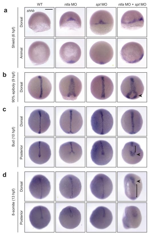
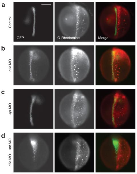
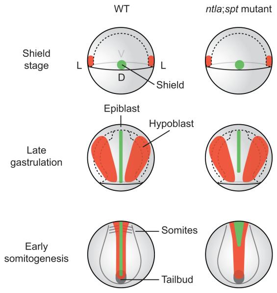
In addition to their cell-autonomous roles in mesoderm development, the zebrafish T-box transcription factors no tail a (ntla) and spadetail (spt/tbx16) are required for medial floor plate (MFP) formation. Posterior MFP cells are completely absent in zebrafish embryos lacking both Ntla and Spt function, and genetic mosaic analyses have shown that the two T-box genes promote MFP development in a non-cell-autonomous manner. On the basis of these observations, it has been proposed that Ntla/Spt-dependent mesoderm-derived signals are required for the induction of posterior but not anterior MFP cells. To investigate the mechanisms by which Ntla and Spt regulate MFP development, we have used photoactivatable caged morpholinos (cMOs) to silence these T-box genes with spatiotemporal control. We find that posterior MFP formation requires Ntla or Spt activity during early gastrulation, specifically in lateral margin-derived cells that converge toward the midline during epiboly and somitogenesis. Nodal signaling-dependent MFP specification is maintained in the absence of Ntla and Spt function; however, midline cells in ntla;spt morphants exhibit aberrant morphogenetic movements, resulting in their anterior mislocalization. Our findings indicate that Ntla and Spt do not differentially regulate MFP induction along the anterior-posterior axis; rather, the T-box genes act redundantly within margin-derived cells to promote the posterior extension of MFP progenitors.
Figures


Similar articles
-
A novel cold-sensitive mutant of ntla reveals temporal roles of brachyury in zebrafish.Dev Dyn. 2016 Aug;245(8):874-80. doi: 10.1002/dvdy.24417. Epub 2016 May 27. Dev Dyn. 2016. PMID: 27153483 Free PMC article.
-
The zebrafish T-box genes no tail and spadetail are required for development of trunk and tail mesoderm and medial floor plate.Development. 2002 Jul;129(14):3311-23. doi: 10.1242/dev.129.14.3311. Development. 2002. PMID: 12091302
-
Spatio-temporal regulation of Wnt and retinoic acid signaling by tbx16/spadetail during zebrafish mesoderm differentiation.BMC Genomics. 2010 Sep 9;11:492. doi: 10.1186/1471-2164-11-492. BMC Genomics. 2010. PMID: 20828405 Free PMC article.
-
Teasing out T-box targets in early mesoderm.Curr Opin Genet Dev. 2008 Oct;18(5):418-25. doi: 10.1016/j.gde.2008.07.017. Epub 2008 Sep 7. Curr Opin Genet Dev. 2008. PMID: 18778771 Free PMC article. Review.
-
T-targets: clues to understanding the functions of T-box proteins.Dev Growth Differ. 2001 Feb;43(1):1-11. doi: 10.1046/j.1440-169x.2001.00556.x. Dev Growth Differ. 2001. PMID: 11148447 Review.
Cited by
-
Targeted cell ablation in zebrafish using optogenetic transcriptional control.Development. 2020 Jun 17;147(12):dev183640. doi: 10.1242/dev.183640. Development. 2020. PMID: 32414936 Free PMC article.
-
tbx6l and tbx16 are redundantly required for posterior paraxial mesoderm formation during zebrafish embryogenesis.Dev Dyn. 2017 Oct;246(10):759-769. doi: 10.1002/dvdy.24547. Epub 2017 Aug 30. Dev Dyn. 2017. PMID: 28691257 Free PMC article.
-
The Development and Application of Opto-Chemical Tools in the Zebrafish.Molecules. 2022 Sep 22;27(19):6231. doi: 10.3390/molecules27196231. Molecules. 2022. PMID: 36234767 Free PMC article. Review.
-
Combinatorial control of gene function with wavelength-selective caged morpholinos.Methods Enzymol. 2019;624:69-88. doi: 10.1016/bs.mie.2019.04.007. Epub 2019 Apr 25. Methods Enzymol. 2019. PMID: 31370936 Free PMC article.
-
Illuminating developmental biology through photochemistry.Nat Chem Biol. 2017 May 17;13(6):587-598. doi: 10.1038/nchembio.2369. Nat Chem Biol. 2017. PMID: 28514427 Free PMC article.
References
-
- Placzek M, Briscoe J. The floor plate: multiple cells, multiple signals. Nat. Rev. Neurosci. 2005;6:230–240. - PubMed
-
- Placzek M, Yamada T, Tessier-Lavigne M, Jessell T, Dodd J. Control of dorsoventral pattern in vertebrate neural development: induction and polarizing properties of the floor plate. Development. 1991;(Suppl 2):105–122. - PubMed
-
- Echelard Y, Epstein DJ, St-Jacques B, Shen L, Mohler J, McMahon JA, McMahon AP. Sonic hedgehog, a member of a family of putative signaling molecules, is implicated in the regulation of CNS polarity. Cell. 1993;75:1417–1430. - PubMed
-
- Kennedy TE, Serafini T, de la Torre JR, Tessier-Lavigne M. Netrins are diffusible chemotropic factors for commissural axons in the embryonic spinal cord. Cell. 1994;78:425–435. - PubMed
-
- Serafini T, Kennedy TE, Galko MJ, Mirzayan C, Jessell TM, Tessier-Lavigne M. The netrins define a family of axon outgrowth-promoting proteins homologous to C. elegans UNC-6. Cell. 1994;78:409–424. - PubMed
Publication types
MeSH terms
Substances
Grants and funding
LinkOut - more resources
Full Text Sources
Other Literature Sources
Molecular Biology Databases
