

Glycation of nail proteins: from basic biochemical findings to a representative marker for diabetic glycation-associated target organ damage
- PMID: 25781337
- PMCID: PMC4363324
- DOI: 10.1371/journal.pone.0120112
Glycation of nail proteins: from basic biochemical findings to a representative marker for diabetic glycation-associated target organ damage
Abstract
Background: Although assessment of glycated nail proteins may be a useful marker for monitoring of diabetes, their nature and formation are still poorly understood. Besides a detailed anatomical analysis of keratin glycation, the usefulness of glycated nail protein assessment for monitoring diabetic complications was investigated.
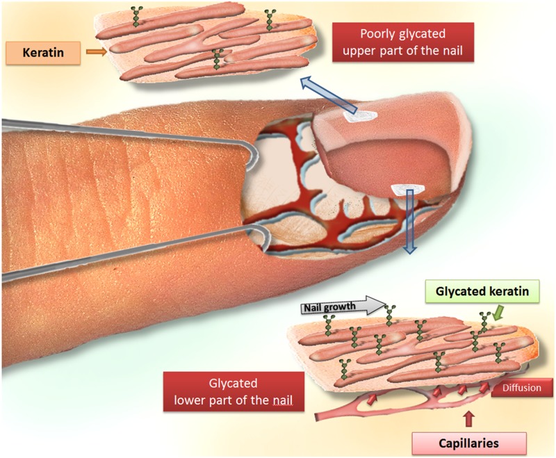
Methods: 216 patients (94 males, 122 females; mean age ± standard deviation: 75.0 ± 8.7 years) were enrolled. Glycation of nail and eye lens proteins was assessed using a photometric nitroblue tetrazolium-based assay. Following chromatographic separation of extracted nail proteins, binding and nonbinding fractions were analyzed using one-dimensional gel electrophoresis. Using a hand piece containing a latch-type-bur, a meticulous cutting of the nail plate into superficial and deep layers was performed, followed by a differential analysis of fructosamine.
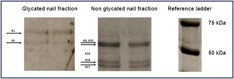
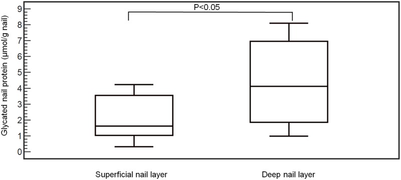
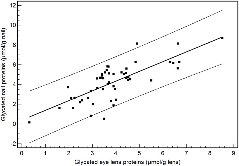
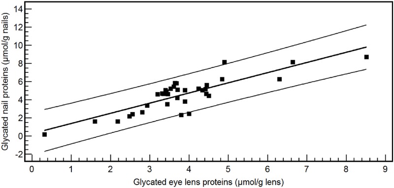
Results: Using SDS PAGE, four and two bands were identified among the nonglycated and glycated nail fraction respectively. Significantly lower fructosamine concentrations were found in the superficial nail layer (mean: 2.16 ± 1.37 μmol/g nails) in comparison with the deep layer (mean: 4.36 ± 2.55 μmol/g nails) (P<0.05). A significant higher amount of glycated eye lens proteins was found in diabetes mellitus patients (mean: 3.80 ± 1.57 μmol/g eye lens) in comparison with nondiabetics (mean: 3.35 ± 1.34 μmol/g eye lens) (P<0.05). A marked correlation was found between glycated nail and glycated eye lens proteins [y (glycated nail proteins) = 0.39 + 0.99 x (eye lens glycated proteins); r2 = 0.58, P<0.001]. The concentration of glycated eye lens proteins and the HbA1c level were found to be predictors of the concentration of glycated nail proteins.
Conclusions: Glycation of nail proteins takes place in the deep layer of finger nails, which is in close contact with blood vessels and interstitial fluid. Glycation of nail proteins can be regarded as a representative marker for diabetic glycation-associated target organ damage.
Conflict of interest statement
Figures
Similar articles
-
Immunochemical detection of glycated beta- and gamma-crystallins in lens and their circulating autoantibodies (IgG) in streptozocin induced diabetic rat.Mol Vis. 2006 Sep 13;12:1077-85. Mol Vis. 2006. PMID: 17093392
-
Glycation and advanced glycation end-products in laboratory experiments in vivo and in vitro.Acta Medica (Hradec Kralove). 2006;49(1):35-9. Acta Medica (Hradec Kralove). 2006. PMID: 16696441
-
Assessment of Absorption of Glycated Nail Proteins in Patients with Diabetes Mellitus and Diabetic Retinopathy.Medicina (Kaunas). 2020 Nov 29;56(12):658. doi: 10.3390/medicina56120658. Medicina (Kaunas). 2020. PMID: 33260342 Free PMC article.
-
The contribution of glycation to cataract formation in diabetes.J Am Optom Assoc. 1998 Aug;69(8):519-30. J Am Optom Assoc. 1998. PMID: 9747048 Review.
-
[Role of nonenzymatic glycation and oxidative stress on the development of complicated diabetic cataracts].Cesk Fysiol. 2000 Feb;49(1):16-21. Cesk Fysiol. 2000. PMID: 10953502 Review. Czech.
Cited by
-
Predicting and preventing diabetes: Translational potential of Ayurveda information on pre-diabetes.J Ayurveda Integr Med. 2021 Oct-Dec;12(4):733-738. doi: 10.1016/j.jaim.2021.05.009. Epub 2021 Jul 16. J Ayurveda Integr Med. 2021. PMID: 34275702 Free PMC article.
-
Use of a Miniaturized Near-Infrared Spectroscopy Device for Type 2 Diabetes Mellitus Screening: Pooled Analysis of the Pilot ANODE01 and ANODE02 Studies.J Diabetes Sci Technol. 2025 Apr 12:19322968251331069. doi: 10.1177/19322968251331069. Online ahead of print. J Diabetes Sci Technol. 2025. PMID: 40219802 Free PMC article.
-
On the nature of toenail opacities in renal insufficiency.Clin Exp Nephrol. 2019 Jan;23(1):146-147. doi: 10.1007/s10157-018-1576-0. Epub 2018 May 5. Clin Exp Nephrol. 2019. PMID: 29730728 No abstract available.
-
Can fingernail quality predict bone damage in Type 2 diabetes mellitus? a pilot study.PLoS One. 2021 Sep 30;16(9):e0257955. doi: 10.1371/journal.pone.0257955. eCollection 2021. PLoS One. 2021. PMID: 34591909 Free PMC article.
-
Glycated nail proteins as a new biomarker in management of the South Kivu Congolese diabetics.Biochem Med (Zagreb). 2015 Oct 15;25(3):469-73. doi: 10.11613/BM.2015.048. eCollection 2015. Biochem Med (Zagreb). 2015. PMID: 26526975 Free PMC article.
References
-
- Nawale RB, Mourya VK, Bhise SB (2006) Non-enzymatic glycation of proteins: a cause for complications in diabetes. Indian J Biochem Biophys 43: 337–344. - PubMed
-
- Horvat S, Jakas A (2004) Peptide and amino acid glycation: new insights into the Maillard reaction. J Pept Sci 10: 119–137. - PubMed
-
- Brownlee M (1992) Glycation products and the pathogenesis of diabetic complications. Diabetes Care 15: 1835–1843. - PubMed
-
- Tessier FJ (2010) The Maillard reaction in the human body. The main discoveries and factors that affect glycation. Pathol Biol (Paris) 58: 214–219. - PubMed
Publication types
MeSH terms
Substances
LinkOut - more resources
Full Text Sources
Other Literature Sources
Medical
