Review
. 2015 Jan;21(1):89-106.
doi: 10.1515/hmbci-2014-0049.

Molecular cues on obesity signals, tumor markers and endometrial cancer

Review

Molecular cues on obesity signals, tumor markers and endometrial cancer

Danielle Daley-Brown et al. Horm Mol Biol Clin Investig. 2015 Jan.

Abstract

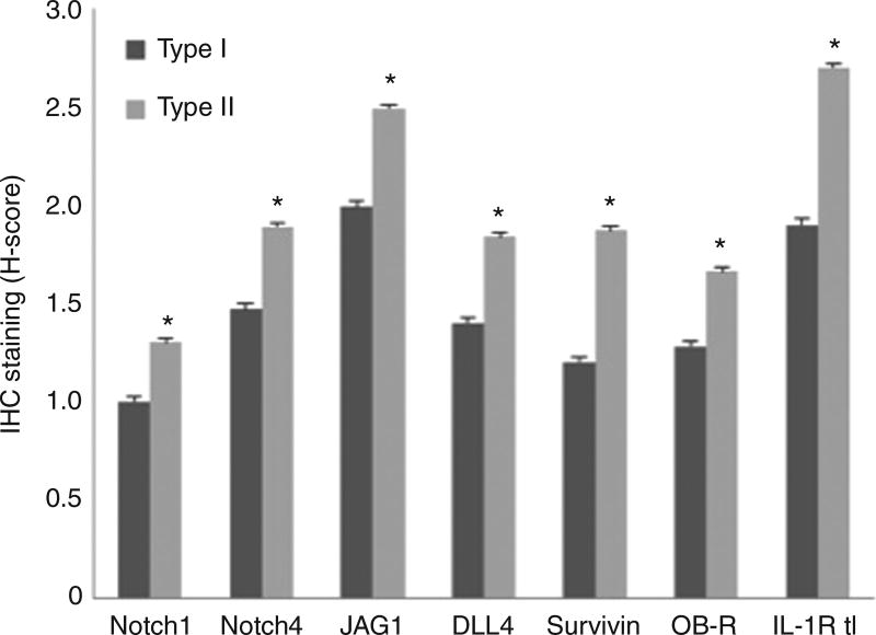
Tumor markers are important tools for early diagnosis, prognosis, therapy response and endometrial cancer monitoring. A large number of molecular and pathologic markers have been described in types I and II endometrial cancers, which has served to define the main oncogenic, epidemiological, genetic, clinical and histopathological features. Ongoing attempts to stratify biological markers of endometrial cancer are presented. However, data on changes in tumor marker profiles in obesity-related endometrial cancer are scarce. Obesity is a pandemic in Western countries that has an important impact on endometrial cancers, albeit through not very well-defined mechanisms. Although endometrial cancer is more common in Caucasian women, higher mortality is found in African Americans who also show higher incidence of obesity. Here, we describe how obesity signals (estrogen, leptin, leptin induced-molecules, Notch; cytokines and growth factors) could affect endometrial cancer. Leptin signaling and its crosstalk may be associated to the more aggressive and poor prognosis type II endometrial cancer, which affects more postmenopausal and African-American women. In this regard, studies on expression of novel molecular markers (Notch, interleukin-1 and leptin crosstalk outcome) may provide essential clues for detection, prevention, treatment and prognosis.


Conflict of interest statement

The authors declare that they have no conflict of interests.

Figures

Figure 1
Figure 1. Type I EmCa (A, B) and type II EmCa (C, D)
(A) Endometrioid carcinoma containing glandular features that resemble glands of the benign endometrium (HE, 10×). (B) Estrogen receptor positive tumor (HE, 40×). (C) Serous carcinoma of the endometrium (HE, 40×). (D) Endometrial carcinoma in situ (HE, 60×).
Figure 2
Figure 2. H-scores from immunohistochemistry staining of NILCO components and targets in type I and type II EmCa from AAW
H-scores were significantly higher in type II versus type I EmCa for Notch1 (p=0.0044), Notch4 (p=0.0081), JAG1 (p=0.0116), DLL4 (p=0.0042), survivin (p=0.0144), OB-R (p=0.0008) and IL-1R tI (p=0.0028).
Figure 3
Figure 3. EmCa derived from EmCa stem cells (ECSC). (A, B) Osteosarcomas; (C, D) chondrosarcomas; (E) rhabdosarcoma; (F) epithelial sarcomatous and (G) choriocarcinoma differentiation
(A) Sarcomatous stromal cells produce osteoid. Tumoral osteoid represented by amorphous fibrillary eosinophilic deposits. Early osteoid deposition forms a lace-like pattern around tumor cells, and advanced osteoid shows evidence of mineralization (darker pink-purple color) (HE, 10×). (B) High magnification shows highly malignant cells with high nuclear-to-cytoplasmic ratio, anaplastic hyperchromatic nuclei or clearing of the chromation and conspicuous, cherry-red nucleoli (HE, 40×). (C) Sarcomatous stromal cells produce chondroid matrix (HE, 10×). (D) Malignant nuclear anaplastic features (HE, 40×). (E) Anaplastic rhabdomyosarcoma showing large strap cells with abundant cytoplasm and striations. The cells are mononuclear or multinucleated. Numerous mitoses are identified (HE, 40×). (F) Epithelial and sarcomatous component blend in this tumor. The malignant epithelial component is composed of cells with high nuclear-to-cytoplasm ratios that show numerous mitoses. Spindle cells with large anaplastic nuclei and prominent nucleoli representing the sarcomatous component are immediately adjacent to the epithelial component (HE, 40×). (G) Malignant polygonal/round cells with single nucleus are reminiscent of the cytotrophoblast. Few multinucleated cells reminiscent of the syncytiotrophoblast are also present. Hemorrhagic background and necrosis areas are present in this tumor (HE, 40×).

References

    1. NIH/NCI website. 2014 http://www.cancer.gov.
    1. Amant F, Moerman P, Neven P, Timmerman D, Van Limbergen E, Vergote I. Endometrial cancer. Lancet. 2005;366:491–505. - PubMed
    1. American Cancer Society, Statistics, 2014. 2014 http://www.cancer.org/research/cancerfactsstatistics/cancerfactsfigures.
    1. Long B, Liu FW, Bristow RE. Disparities in uterine cancer epidemiology, treatment, and survival among African Americans in the United States. Gynecol Oncol. 2013;130:652–9. - PMC - PubMed
    1. Schmandt RE, Iglesias DA, Co NN, Lu KH. Understanding obesity and endometrial cancer risk: opportunities for prevention. Am J Obstet Gynecol. 2011;205:518–25. - PMC - PubMed

Publication types

Substances

LinkOut - more resources