. 2015 Mar 17;10(3):e0120352.
doi: 10.1371/journal.pone.0120352. eCollection 2015.

Assembly and interrogation of Alzheimer's disease genetic networks reveal novel regulators of progression

Affiliations

Assembly and interrogation of Alzheimer's disease genetic networks reveal novel regulators of progression

Soline Aubry et al. PLoS One. .

Abstract

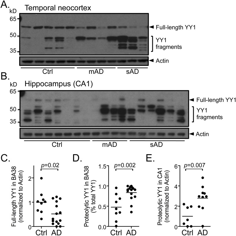
Alzheimer's disease (AD) is a complex multifactorial disorder with poorly characterized pathogenesis. Our understanding of this disease would thus benefit from an approach that addresses this complexity by elucidating the regulatory networks that are dysregulated in the neural compartment of AD patients, across distinct brain regions. Here, we use a Systems Biology (SB) approach, which has been highly successful in the dissection of cancer related phenotypes, to reverse engineer the transcriptional regulation layer of human neuronal cells and interrogate it to infer candidate Master Regulators (MRs) responsible for disease progression. Analysis of gene expression profiles from laser-captured neurons from AD and controls subjects, using the Algorithm for the Reconstruction of Accurate Cellular Networks (ARACNe), yielded an interactome consisting of 488,353 transcription-factor/target interactions. Interrogation of this interactome, using the Master Regulator INference algorithm (MARINa), identified an unbiased set of candidate MRs causally responsible for regulating the transcriptional signature of AD progression. Experimental assays in autopsy-derived human brain tissue showed that three of the top candidate MRs (YY1, p300 and ZMYM3) are indeed biochemically and histopathologically dysregulated in AD brains compared to controls. Our results additionally implicate p53 and loss of acetylation homeostasis in the neurodegenerative process. This study suggests that an integrative, SB approach can be applied to AD and other neurodegenerative diseases, and provide significant novel insight on the disease progression.


Conflict of interest statement

Competing Interests: The authors have declared that no competing interests exist.

Figures

Fig 1
Fig 1. Cluster dendrograms for hippocampus and visual cortex samples.
Bioinformatic analysis reveals a high degree of clustering of Alzheimer disease (AD), non-demented AD (NDAD) and control (C) in the hippocampus but not the primary visual cortex. Dendrogram showing clustering of (A) the hippocampus and (B) primary visual cortex samples using centered correlation and average linkage.
Fig 2
Fig 2. Count of candidate MRs per region and per comparison groups after each computational analysis.
Master Regulator Inference analysis (MARINa) reveals different numbers of candidate Master Regulators (MRs) by brain region and comparison group. Count of candidate MRs in (A) control v. Alzheimer’s disease (AD), (B) control v. non-demented Alzheimer’s disease (NDAD) and (C) non-demented Alzheimer’s disease (NDAD) v. Alzheimer’s disease (AD) after each analysis. EC = entorhinal cortex, HIP = hippocampus, MTG = middle temporal gyrus, PC = posterior cingulate, SFG = superior frontal gyrus, VCX = visual cortex.
Fig 3
Fig 3. Increased proteolysis of YY1 is observed in post-mortem Alzheimer’s disease (AD) brain tissue.
(A, B) Representative immunoblots using nuclear fractions from 12 human brain tissue samples from temporal neocortex (BA38) and 17 from hippocampus (CA1) show nuclear full-length and proteolytic fragments of YY1 in control (Ctrl), moderate Alzheimer’s disease (mAD) and severe Alzheimer’s disease (sAD) cases. Densitometric analysis of immunoblots from BA38 shows (C) a significant decrease (p = 0.02) in the full-length YY1 protein (60 kDa) in AD (n = 15, mAD + sAD) and (D) a significant increase (p = 0.002) in the proportion of proteolytic fragments (35–50 kDa) compared to controls (n = 9). Densitometric analysis of immunoblots from CA1 also reveals a significant increase (p = 0.007) in YY1 proteolytic fragments in AD (n = 10, mAD + sAD) compared to controls (n = 7). Comparisons were made using a Student’s t-test.
Fig 4
Fig 4. Exogenous treatment of rat primary hippocampal cultures with cytotoxic concentrations of dodecomeric preparations of Aβ (Aβdod) results in increased YY1 protein levels and proteolysis.
(A) Western blot analyses of extracts from cells treated with Aβdod for 6 hr show an increase in full-length YY1 as well as the appearance of proteolytic cleavage fragments between 35 and 50 kDa at 10 μM but not 300 nM. (B) Densitometric analysis of the full-length 65 kDa YY1 band shows a statistically significant increase in full-length YY1 in cells treated with 10 μM Aβdod compared to control (p = 0.04). Comparisons were made using a Student’s t-test. Veh = vehicle, tub = tubulin.
Fig 5
Fig 5. Increase in p300 acetyltransferase activity in moderate (mAD) and severe AD (sAD) cases.
Immunohistochemistry and immunofluorescence analyses of phosphor-Ser1834-p300 (p-p300) on paraffin sections from the hippocampal area CA1 of human brains from control (Ctrl) (A, D), moderate AD (mAD) (B, E) and severe AD (sAD) (C, F) show cytoplasmic granular labeling resembling granulovacuolar degeneration in pyramidal neurons (B, C, E, F; arrows) in AD tissue, which co-localize with phospho-tau (p-tau) in ~95% of p-p300-positive neurons (E, F; arrows). p-p300-positive threads (A, D, arrowheads) are also observed in affected tissue. Controls are negative for p-tau and p-p300 (A, D). (G) Immunoblots of nuclear and cytoplasmic fractions of temporal neocortex (BA38) from Ctrl, mAD and sAD cases show an increase in Ac-Lys382-p53 (Ac-p53) in AD (n = 7, mAD+sAD) compared to control (n = 5) in both fractions. Quantification using densitometric analysis reveals a significant increase in Ac-p53 in nuclear (H, p = 0.002) and cytoplasmic (I, p = 0.001) fractions compared to controls. (J) Immunoblots of whole tissue extracts from BA38 from Ctrl, mAD and sAD cases also show a significant increase in Ac-Lys14-Histone 3 (Ac-H3) in mAD (n = 7) and sAD (n = 6) compared to Ctrl (n = 7). (K) Quantification reveals a statistically significant increase in Ac-H3 in mAD (p = 0.004) and sAD (p = 0.0003) compared to control. Comparisons were made using a Student’s t-test. Control case 4 is a statistical outlier, more than 3 standard deviations above the mean, and was excluded from the analysis. (AC) Scale bar = 50 μm, (DF) scale bar = 50 μm.
Fig 6
Fig 6. ZMYM3 protein level significantly decreases in severe AD cases.
(A, B) Representative immunoblots using nuclear fractions from temporal neocortex (BA38) (A) and hippocampal area CA1 (B) show a decrease in the levels of ZMYM3 in severe Alzheimer’s disease (sAD) compared to control (Ctrl) and moderate Alzheimer’s disease (mAD). (C, D) Densitometric analyses of the 70 kDa band shows a significant decrease in ZMYM3 in severe AD cases in BA38 (n = 8; p = 0.0009) and CA1 (n = 6; p = 0.004) compared to controls in BA38 (n = 9) and CA1 (n = 7). Comparisons were made using a Student’s t-test.

References

    1. WHO (2011) Global Health and Aging. World Health Organization.
    1. Selkoe DJ (1991) The molecular pathology of Alzheimer's disease. Neuron 6: 487–498. - PubMed
    1. Thinakaran G, Koo EH (2008) Amyloid precursor protein trafficking, processing, and function. J Biol Chem 283: 29615–29619. 10.1074/jbc.R800019200 - DOI - PMC - PubMed
    1. Wilson CA, Doms RW, Lee VM (1999) Intracellular APP processing and A beta production in Alzheimer disease. J Neuropathol Exp Neurol 58: 787–794. - PubMed
    1. Lefort R, Pozueta J, Shelanski M (2012) Cross-linking of cell surface amyloid precursor protein leads to increased beta-amyloid peptide production in hippocampal neurons: implications for Alzheimer's disease. J Neurosci 32: 10674–10685. 10.1523/JNEUROSCI.6473-11.2012 - DOI - PMC - PubMed

Publication types

Substances