Comparative Study
. 2015 Mar 17;10(3):e0122014.
doi: 10.1371/journal.pone.0122014. eCollection 2015.

Differential regulation of proton-sensitive ion channels by phospholipids: a comparative study between ASICs and TRPV1

Affiliations
Comparative Study

Differential regulation of proton-sensitive ion channels by phospholipids: a comparative study between ASICs and TRPV1

Hae-Jin Kweon et al. PLoS One. .

Abstract

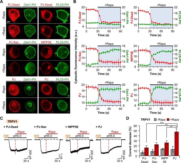
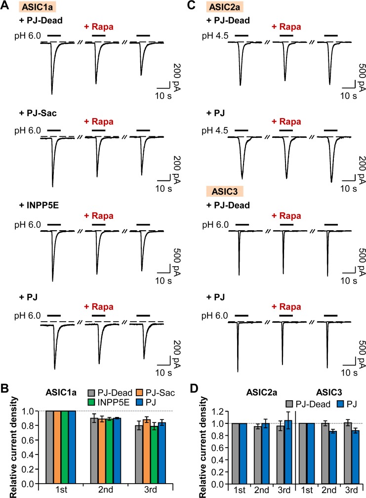
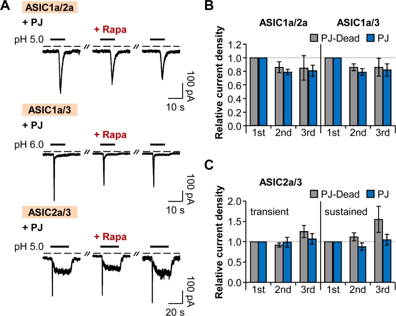
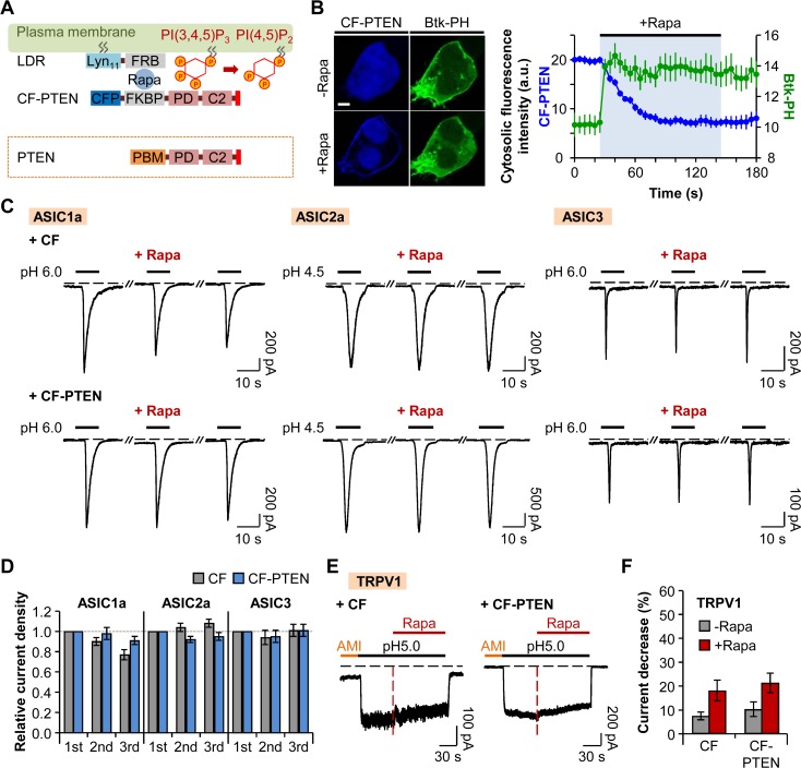
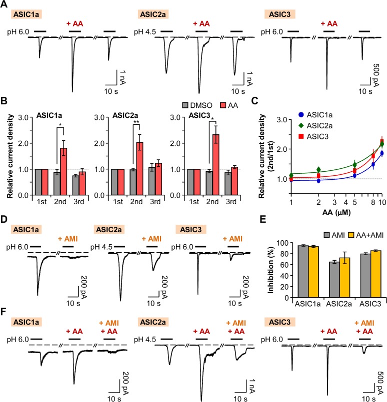
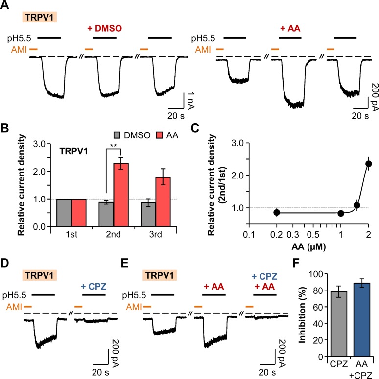
Protons are released in pain-generating pathological conditions such as inflammation, ischemic stroke, infection, and cancer. During normal synaptic activities, protons are thought to play a role in neurotransmission processes. Acid-sensing ion channels (ASICs) are typical proton sensors in the central nervous system (CNS) and the peripheral nervous system (PNS). In addition to ASICs, capsaicin- and heat-activated transient receptor potential vanilloid 1 (TRPV1) channels can also mediate proton-mediated pain signaling. In spite of their importance in perception of pH fluctuations, the regulatory mechanisms of these proton-sensitive ion channels still need to be further investigated. Here, we compared regulation of ASICs and TRPV1 by membrane phosphoinositides, which are general cofactors of many receptors and ion channels. We observed that ASICs do not require membrane phosphatidylinositol 4-phosphate (PI(4)P) or phosphatidylinositol 4,5-bisphosphate (PI(4,5)P2) for their function. However, TRPV1 currents were inhibited by simultaneous breakdown of PI(4)P and PI(4,5)P2. By using a novel chimeric protein, CF-PTEN, that can specifically dephosphorylate at the D3 position of phosphatidylinositol 3,4,5-trisphosphate (PI(3,4,5)P3), we also observed that neither ASICs nor TRPV1 activities were altered by depletion of PI(3,4,5)P3 in intact cells. Finally, we compared the effects of arachidonic acid (AA) on two proton-sensitive ion channels. We observed that AA potentiates the currents of both ASICs and TRPV1, but that they have different recovery aspects. In conclusion, ASICs and TRPV1 have different sensitivities toward membrane phospholipids, such as PI(4)P, PI(4,5)P2, and AA, although they have common roles as proton sensors. Further investigation about the complementary roles and respective contributions of ASICs and TRPV1 in proton-mediated signaling is necessary.


Conflict of interest statement

Competing Interests: The authors have declared that no competing interests exist.

Figures

Fig 1
Fig 1. The activity of TRPV1 is dependent on phosphoinositides.
(A) Confocal images of cells expressing PJ-Dead (top), PJ-Sac (middle), INPP5E (middle), or PJ (bottom) with LDR and respective biosensors for PI(4)P (Osh1-PH-GFP) or PI(4,5)P2 (PLCδ1-PH-GFP). Images before and after the addition of rapamycin (1 μM) for 60 s (Scale bar, 5 μm). (B) Cytosolic fluorescence intensities of RFP (red) and GFP (green) for the cells in (A). The values of the Y-axis use an arbitrary unit. Cells expressing PJ-Dead (top), PJ-Sac (middle), INPP5E (middle), or PJ (bottom) (n = 3, respectively). (C) TRPV1 currents triggered by prolonged extracellular pH drop to 5.0 for 150 s. Rapamycin (1 μM) was co-applied for 90 s during the acid stimuli. Amiloride (300 μM) was pretreated for 30 s before the pH pulse. Black dashed line indicates the zero current level. Red dashed line indicates the point of rapamycin application. (D) Percentage of current decrease in (C) during 45 s of acidification before (grey) and after (red) rapamycin addition (n = 12 for PJ-Dead; n = 14 for PJ-Sac; n = 12 for INPP5E; n = 10 for PJ). ** P < 0.01 and *** P < 0.001, with two-way ANOVA followed by Bonferroni post-hoc test and one-way ANOVA followed by Bonferroni post-hoc test. Data are mean ± SEM.
Fig 2
Fig 2. Homomeric ASIC currents are insensitive to PI(4)P and PI(4,5)P2.
(A) ASIC1a currents evoked by repetitive rapid extracellular pH change from 7.4 to 6.0 for 10 s with time intervals of 120 s in cells expressing LDR and either PJ-Dead, PJ-Sac, INPP5E, or PJ. Rapamycin (1 μM) was bath-applied for 60 s, and then normal extracellular solution was perfused for 10 s right before the second pulse to minimize possible side effects of rapamycin. Dashed line indicates the zero current level. (B) Relative current density measured for the cells in (A) (n = 6, respectively). Current density of each pulse was divided by that of the first pulse. There is no statistical significance with two-way ANOVA followed by Bonferroni post-hoc test. (C) ASIC2a or ASIC3 current traces evoked by pH drop to 4.5 or 6.0 for 10 s. (D) Relative current density measured for the cells in (C) (n = 6, respectively). There is no statistical significance with two-way ANOVA followed by Bonferroni post-hoc test. Data are mean ± SEM.
Fig 3
Fig 3. Heteromeric ASIC currents are insensitive to PI(4)P and PI(4,5)P2.
(A) Current traces from ASIC1a/2a, ASIC1a/3, and ASIC2a/3 heteromeric channels evoked by extracellular acidification in cells expressing LDR and PJ. Rapamycin (1 μM) was bath-applied for 60 s, and then normal extracellular solution was perfused for 10 s right before the second pulse to minimize possible side effects of rapamycin. Dashed line indicates the zero current level. (B) Relative current density measured for the currents of ASIC1a/2a and ASIC1a/3 in (A) (n = 3, respectively). Current density of each pulse was divided by that of the first pulse. There is no statistical significance with two-way ANOVA followed by Bonferroni post-hoc test. (C) Relative current density measured for the transient and sustained currents of ASIC2a/3 in (A) (n = 3 for PJ-Dead; n = 5 for PJ). There is no statistical significance with two-way ANOVA followed by Bonferroni post-hoc test. Data are mean ± SEM.
Fig 4
Fig 4. Neither ASICs nor TRPV1 activities are affected by depletion of PI(3,4,5)P3.
(A) CF-PTEN is rapidly recruited to the plasma membrane anchor LDR by dimerization of FRB and FKBP upon addition of rapamycin, and the PD domain of CF-PTEN specifically dephosphorylates PI(3,4,5)P3 to PI(4,5)P2. Red bar in the C-terminal tail of PTEN indicates PDZ-binding domain. (B) Confocal images of cells expressing LDR, CF-PTEN, and Btk-PH-GFP before and after the addition of rapamycin (1 μM) for 120 s (Scale bar, 5 μm) and cytosolic fluorescence intensities of CFP (blue) and GFP (green) (n = 3). The values of the Y-axis use an arbitrary unit. (C) ASIC current traces triggered by extracellular acidification in cells expressing LDR and CF (lacking PTEN) or CF-PTEN. Rapamycin (1 μM) was bath-applied for 60 s, and then normal extracellular solution was perfused for 10 s right before the second pulse to minimize possible side effects of rapamycin. Dashed line indicates the zero current level. (D) Relative current density measured for the cells in (C) (CF (n = 8), CF-PTEN (n = 7) for ASIC1a; CF (n = 8), CF-PTEN (n = 8) for ASIC2a; and CF (n = 10), CF-PTEN (n = 10) for ASIC3). Current density of each pulse was divided by that of the first pulse. There is no statistical significance with two-way ANOVA followed by Bonferroni post-hoc test. (E) TRPV1 currents in response to pH drop in the cells expressing LDR and CF or CF-PTEN. Rapamycin (1 μM) was co-applied for 90 s during the acid stimuli. Amiloride (300 μM) was pretreated for 30 s before the pH pulse. Black dashed line indicates the zero current level. Red dashed line indicates the point of rapamycin application. (F) Percentage of current decrease in (E) during 45 s of acidification before (grey) and after (red) rapamycin addition (n = 9 for CF; n = 10 for CF-PTEN). Data are mean ± SEM.
Fig 5
Fig 5. Potentiation of ASICs by AA.
(A) ASIC current traces activated by rapid extracellular pH changes. AA (10 μM) was bath-applied for 20 s before the second pulse. Dashed line indicates the zero current level. (B) Relative current density was measured for the cells expressing ASIC1a (n = 5 for DMSO; n = 6 for AA), ASIC2a (n = 5 for DMSO; n = 10 for AA), and ASIC3 (n = 5 for DMSO; n = 12 for AA). Current density of each pulse was divided by that of the first pulse. * P < 0.05 and ** P < 0.01, with two-way ANOVA followed by Bonferroni post-hoc test and student’s t-test. (C) Dose-dependent relative current density of ASIC1a (blue) (n = 5–20), ASIC2a (green) (n = 5–25), and ASIC3 (red) (n = 5–23). (D) ASIC1a and ASIC3 currents were inhibited by preincubation of cells with pH 7.4 solution containing amiloride (300 μM) for 20 s before the second pulse. In the case of ASIC2a, 600 μM of amiloride was applied for 30 s before and during the second pulse. (E) Percentage of inhibition by amiloride in the absence (grey) or the presence (yellow) of AA (AMI (n = 7) and AA+AMI (n = 4) for ASIC1a; AMI (n = 4) and AA+AMI (n = 3) for ASIC2a; and AMI (n = 6) and AA+AMI (n = 6) for ASIC3). (F) The potentiating effect of AA (10 μM) on ASIC currents was inhibited by amiloride. Data are mean ± SEM.
Fig 6
Fig 6. Potentiation of TRPV1 by AA.
(A) TRPV1 current traces repetitively activated by extracellular pH drop to 5.5 for 30 s with time intervals of 300 s. Amiloride (300 μM) was pretreated for 10 s before the pH pulses. AA (2 μM) was applied for 20 s before the second amiloride treatment. Dashed line indicates the zero current level. (B) Relative current density was measured for the cells in (A) (n = 4 for DMSO; n = 9 for AA). Current density of each pulse was divided by that of the first pulse. ** P < 0.01, with two-way ANOVA followed by Bonferroni post-hoc test and student’s t-test. (C) Dose-dependent relative current density of TRPV1 (n = 5–17). (D) TRPV1 currents were inhibited by preincubation of cells with pH 7.4 solution containing capsazepine (10 μM) for 20 s before the second amiloride treatment. (E) The potentiating effect of AA (2 μM) on TRPV1 currents was inhibited by capsazepine. (F) Percentage of inhibition by capsazepine in the absence (grey) or the presence (blue) of AA (n = 6 for CPZ; n = 6 for AA+CPZ). Data are mean ± SEM.

Similar articles

Cited by

References

    1. Kweon HJ, Suh BC. Acid-sensing ion channels (ASICs): therapeutic targets for neurological diseases and their regulation. BMB Rep. 2013;46(6): 295–304. - PMC - PubMed
    1. Du J, Reznikov LR, Price MP, Zha XM, Lu Y, Moninger TO, et al. Protons are a neurotransmitter that regulates synaptic plasticity in the lateral amygdala. Proc Natl Acad Sci. 2014;111(24): 8961–6. 10.1073/pnas.1407018111 - DOI - PMC - PubMed
    1. Deval E, Gasull X, Noël J, Salinas M, Baron A, Diochot S, et al. Acid-sensing ion channels (ASICs): pharmacology and implication in pain. Pharmacol Ther. 2010;128(3): 549–58. 10.1016/j.pharmthera.2010.08.006 - DOI - PubMed
    1. Smith ML, von Hanwehr R, Siesjö BK. Changes in extra- and intracellular pH in the brain during and following ischemia in hyperglycemic and in moderately hypoglycemic rats. J Cereb Blood Flow Metab. 1986;6(5): 574–83. - PubMed
    1. Magnotta VA, Heo HY, Dlouhy BJ, Dahdaleh NS, Follmer RL, Thedens DR, et al. Detecting activity-evoked pH changes in human brain. Proc Natl Acad Sci. 2012;109(21): 8270–3. 10.1073/pnas.1205902109 - DOI - PMC - PubMed

Publication types