Review
. 2015 Jul 8:38:25-46.
doi: 10.1146/annurev-neuro-071714-033835. Epub 2015 Mar 12.

Neuronal and vascular interactions

Affiliations
Review

Neuronal and vascular interactions

Benjamin J Andreone et al. Annu Rev Neurosci. .

Abstract

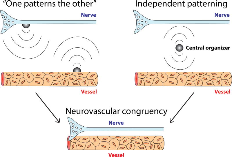
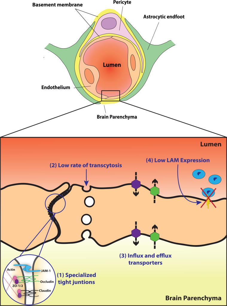
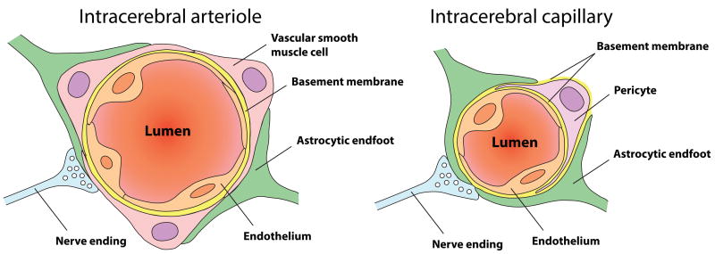
The brain, which represents 2% of body mass but consumes 20% of body energy at rest, has a limited capacity to store energy and is therefore highly dependent on oxygen and glucose supply from the blood stream. Normal functioning of neural circuits thus relies on adequate matching between metabolic needs and blood supply. Moreover, not only does the brain need to be densely vascularized, it also requires a tightly controlled environment free of toxins and pathogens to provide the proper chemical composition for synaptic transmission and neuronal function. In this review, we focus on three major factors that ensure optimal brain perfusion and function: the patterning of vascular networks to efficiently deliver blood and nutrients, the function of the blood-brain barrier to maintain brain homeostasis, and the regulation of cerebral blood flow to adequately couple energy supply to neural function.

Keywords: blood–brain barrier; cerebral blood flow; cerebrovascular patterning; endothelial cells; neurovascular networks; neurovascular unit.


Figures

Figure 1
Figure 1
Two models of Neurovascular congruency: (Left) “One patterns the other” model, in which either the nervous or vascular system precedes developmentally, and then instructs the other system to form using its established architecture as a template. In the case of a tissue with a planar structure such as skin, or during pathfinding before nerves and vessel reach their target tissue, this model allows for the development of parallel trajectories of nerves and vessels, independent of their position relative to their surroundings. (Right) Independent patterning model, in which balanced attractive and repulsive signals originate from a central organizer within a target tissue – this central organizer acts to pattern neurovascular congruency. When nerves and vessels reach a target tissue with a complex three-dimensional structure, the precise architecture of nerves, vessels and the target tissue becomes functionally relevant, as dictated by the unique requirements of the target tissue environment.
Figure 2
Figure 2
CNS endothelial cell properties contributing to BBB functionality: (Top) A cross-sectional view of the neurovascular unit at the level of a CNS capillary. Vessels are lined by a single-layer of non-fenestrated endothelial cells that exhibit barrier properties. Astrocytes and pericytes surround the abluminal surface of CNS endothelial cells and provide additional functional support for the establishment and maintenance of the BBB. (Bottom) A magnified view of the CNS endothelium, highlighting the four cellular properties that contribute to BBB integrity by stringently controlling the exchange of ions and nutrients between the blood and brain. (1) Specialized tight junctions prevent paracellular flux between endothelial cells. Cell-cell interactions are mediated by tight junction proteins, including junctional adhesion molecule-1 (JAM-1), occludin, and members of the claudin family. The cytoplasmic adaptor proteins ZO-1 and ZO-2 link these transmembrane proteins to the cytoskeleton. (2) Endothelial cells exhibit extremely low rates of transcytosis, as vesicular trafficking of ions and nutrients across cells is kept to a minimum. (3) Endothelial cells express influx (purple hexagons) and efflux (green hexagons) transporters that both shuttle specific nutrients into the brain and remove potentially harmful toxins and other small molecules from the brain, respectively. (4) The low expression of leukocyte adhesion molecules (LAM) aids in maintaining low levels of immune surveillance in the CNS.
Figure 3
Figure 3
Schematic representation of the neurovascular unit: Inside the brain, endothelial cells are organized into a multicellular complex together with contractile and glial cells, an assembly called the neurovascular unit. The main difference between intracerebral arterioles and capillaries is the nature, position and abundance of contractile cells that surround the external vessel surface. (Left) At the level of intracerebral arterioles, the endothelium is fully covered by a single layer of vascular smooth muscle cells which provide contractile properties to the arteriole. Astrocytes send their processes called “end-feet” around the arteriole, providing further support as well as a functional connection to surrounding neural tissues. (Right) Intracerebral capillaries lack vascular smooth muscle cells but are partly covered by contractile pericytes, with a higher density in the central nervous system. Recent findings provide new evidence about the importance of pericytes contractile properties in neurovascular coupling.

Similar articles

Cited by

References

    1. Adams RH, Eichmann A. Axon guidance molecules in vascular patterning. Cold Spring Harbor perspectives in biology. 2010;2:a001875. - PMC - PubMed
    1. Aird WC. Phenotypic heterogeneity of the endothelium: I. Structure, function, and mechanisms. Circ Res. 2007;100:158–73. - PubMed
    1. Alvarez JI, Dodelet-Devillers A, Kebir H, Ifergan I, Fabre PJ, et al. The Hedgehog pathway promotes blood-brain barrier integrity and CNS immune quiescence. Science. 2011;334:1727–31. - PubMed
    1. Anderson AW, Marois R, Colson ER, Peterson BS, Duncan CC, et al. Neonatal auditory activation detected by functional magnetic resonance imaging. Magnetic resonance imaging. 2001;19:1–5. - PubMed
    1. Argandona EG, Lafuente JV. Effects of dark-rearing on the vascularization of the developmental rat visual cortex. Brain Res. 1996;732:43–51. - PubMed