Review
. 2015 Mar 17;8(368):re2.
doi: 10.1126/scisignal.2005813.

Outside the coding genome, mammalian microRNAs confer structural and functional complexity

Affiliations
Review

Outside the coding genome, mammalian microRNAs confer structural and functional complexity

Virginie Olive et al. Sci Signal. .

Abstract

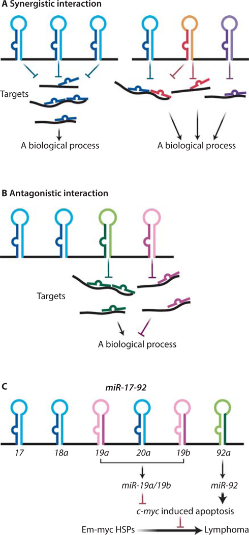
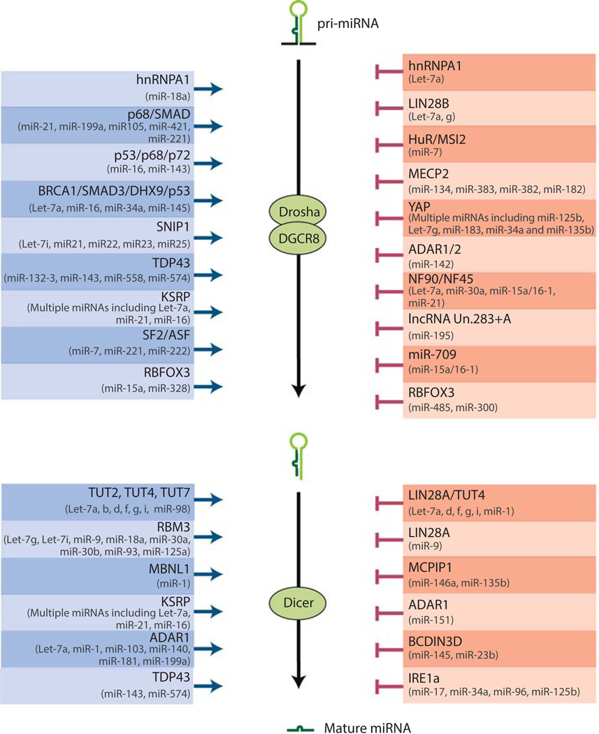
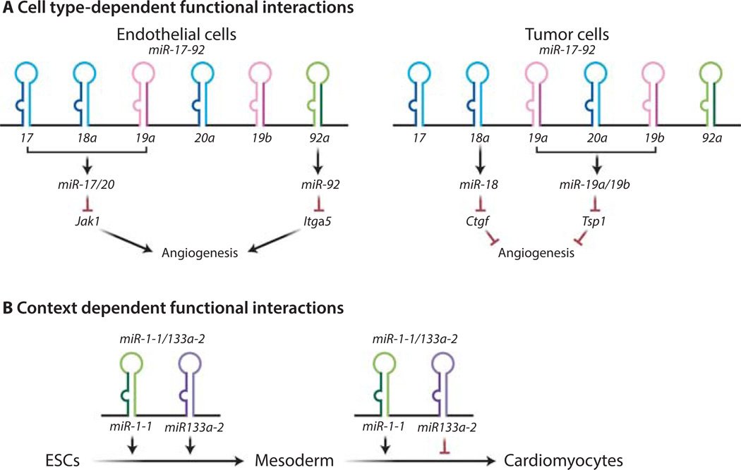
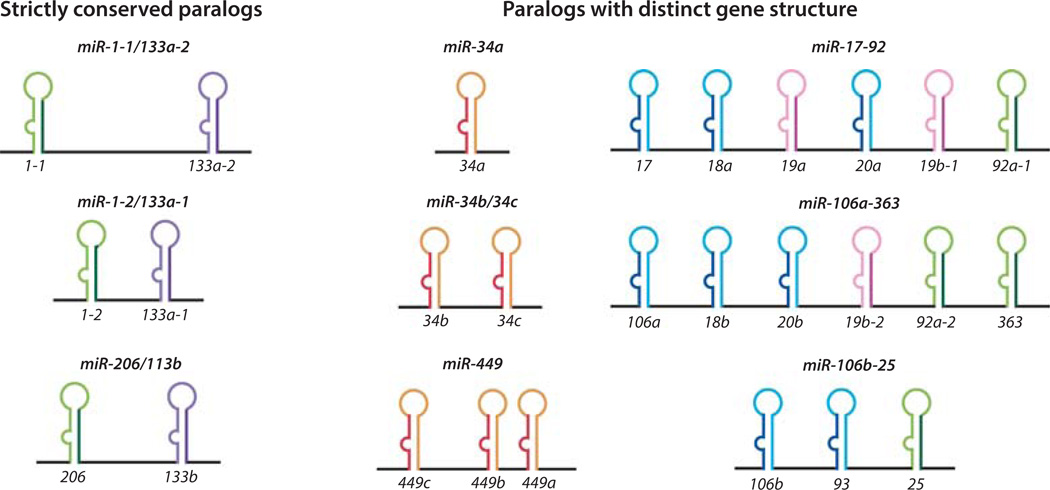
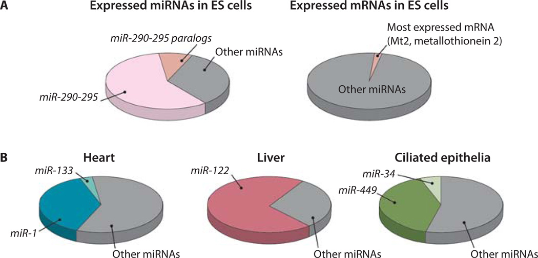
MicroRNAs (miRNAs) comprise a class of small, regulatory noncoding RNAs (ncRNAs) with pivotal roles in posttranscriptional gene regulation. Since their initial discovery in 1993, numerous miRNAs have been identified in mammalian genomes, many of which play important roles in diverse cellular processes in development and disease. These small ncRNAs regulate the expression of many protein-coding genes posttranscriptionally, thus adding a substantial complexity to the molecular networks underlying physiological development and disease. In part, this complexity arises from the distinct gene structures, the extensive genomic redundancy, and the complex regulation of the expression and biogenesis of miRNAs. These characteristics contribute to the functional robustness and versatility of miRNAs and provide important clues to the functional significance of these small ncRNAs. The unique structure and function of miRNAs will continue to inspire many to explore the vast noncoding genome and to elucidate the molecular basis for the functional complexity of mammalian genomes.


Figures

Figure 1
Figure 1. Polycistronic miRNAs harbor complex functional interactions among its components
(A) Cooperative functional interaction occurs within a polycistronic miRNA. Some polycistronic miRNAs contain a tandem of homologous components that frequently share the same seed sequences, thus they often have the same targets and biological function (right panel). Other miRNA clusters consist of heterologous miRNAs that act on different target sites to synergically regulate one biological process (left panel). (B) miRNA polycistrons can harbor a functional antagonism among the encoded components. (C) A schematic illustration of the miR-19:miR-92 antagonism in regulating the oncogenic cooperation between miR-17–92 and c-Myc. Whereas miR-19 miRNAs repressed c-Myc-induced apoptosis to promote oncogenesis, miR-92 exhibits an opposite effect to promote c-Myc induced apoptosis.
Figure 2
Figure 2. Diverse mechanisms regulate miRNA-specific biogenesis
A schematic illustration summarizes the biogenesis regulators of specific miRNAs, with positive regulators on the left and negative regulators on the right (, , , , , , , , , , , , , , , –, , –158, 88, 95, 159, 160). miRNAs whose biogenesis is controlled by each regulator also are listed.
Figure 3
Figure 3. Cell type- and context-dependent versatility of polycistronic miRNA functions
(A) The miR-17–92 functions on angiogenesis depend on cell types. miR-18a and miR-19 repress the abundance of several secreted molecules in tumor cells (54), thereby promoting angiogenesis through a cell non-autonomous mechanism. In contrast, in endothelial cells miR-17/20 and miR-92a suppress the abundance of JAK and the integrin subunit α5, respectively, thus inducing an anti-angiogenic effect through a cell autonomous mechanism (55, 56). (B) The mode of functional interaction between miR-1 and miR-133 depends on the biological contexts. Although miR-1 and miR-133 synergistically promote differentiation from embryonic stem cells to mesoderm, they exhibit an functional antagonism in regulating the muscle-specific differentiation from mesoderm (58).
Figure 4
Figure 4. Distinct genomic organization of paralogous polycistronic miRNA loci
miR-1-1/133a-2, miR-1-2/133a-1 and miR-206/133b are strictly conserved paralogs. miR-34a, miR-34b/34c and miR-449 are paralogs with distinct genes structure. They contain homologous miRNAs that are present in different copy numbers. miR-17–92, miR-106a-363 and miR-106b-25 are also paralogs that harbor distinct gene structures. They are composed of both homologous and heterologous miRNAs, and only a subset of components is conserved between the paralogs.
Figure 5
Figure 5. The dominant expression of miRNAs in specific cell types
(A) The distribution of the most abundant miRNA and mRNA in the transcriptome. miR-290–295 and its homologs constitute ~70% of total expressed miRNAs in ES cells (81). In contrast, the most abundant mRNA in ES cells constitutes 1.3% of the total transcriptome (Risso et al., personal communication). (B) The dominantly expressed miRNA families in heart tissue, hepatocytes and ciliated epithelia (GEO :GSM539871) (84, 85).

References

    1. Mattick JS. Legislative Activity: RNA regulation: a new genetics? 2004;5:1662–1666.
    1. Djebali S, et al. Landscape of transcription in human cells. Nature. 2012;489:101–108. - PMC - PubMed
    1. Gerstein MB, et al. Architecture of the human regulatory network derived from ENCODE data. Nature. 2012;489:91–100. - PMC - PubMed
    1. L Rinn J, Chang HY. Genome regulation by long noncoding RNAs. Annu. Rev. Biochem. 2012;81:145–166. - PMC - PubMed
    1. Cech TR, Steitz JA. The noncoding RNA revolution-trashing old rules to forge new ones. Cell. 2014;157:77–94. - PubMed

Publication types