

The ureteric bud epithelium: morphogenesis and roles in metanephric kidney patterning
- PMID: 25783232
- PMCID: PMC4376585
- DOI: 10.1002/mrd.22462
The ureteric bud epithelium: morphogenesis and roles in metanephric kidney patterning
Abstract
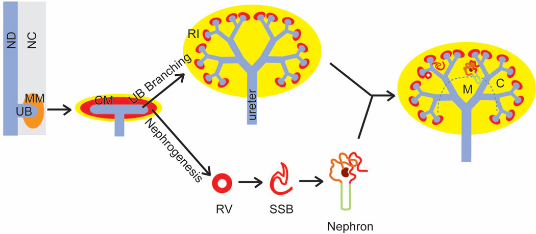
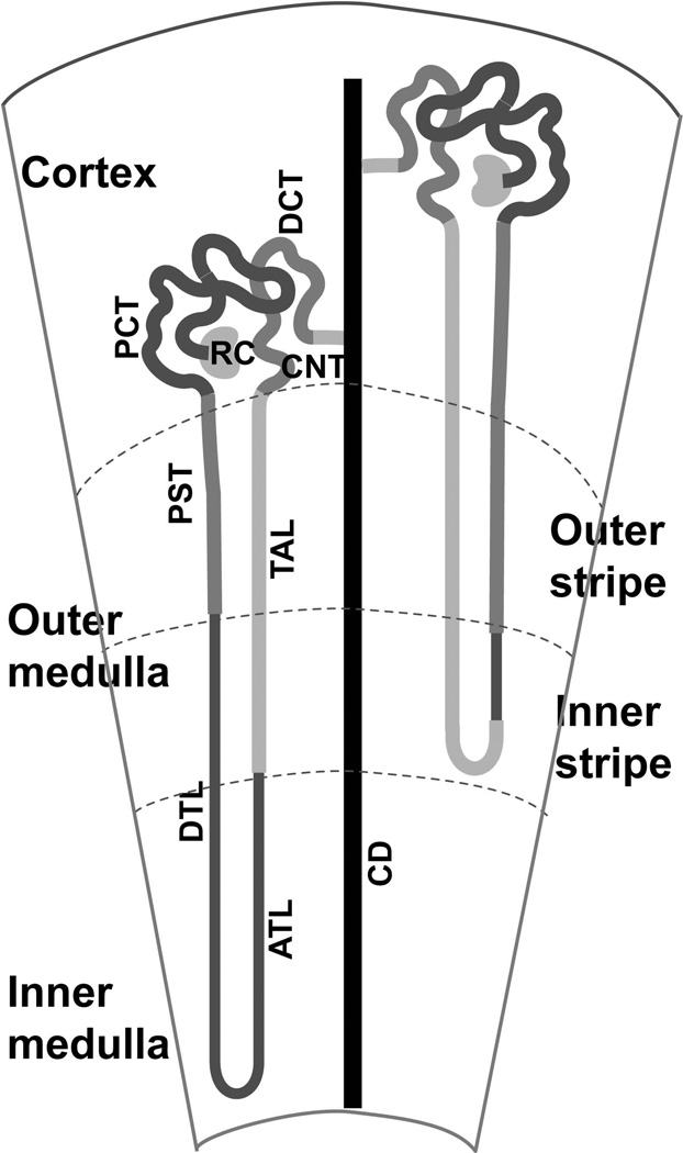
The mammalian metanephric kidney is composed of two epithelial components, the collecting duct system and the nephron epithelium, that differentiate from two different tissues -the ureteric bud epithelium and the nephron progenitors, respectively-of intermediate mesoderm origin. The collecting duct system is generated through reiterative ureteric bud branching morphogenesis, whereas the nephron epithelium is formed in a process termed nephrogenesis, which is initiated with the mesenchymal-epithelial transition of the nephron progenitors. Ureteric bud branching morphogenesis is regulated by nephron progenitors, and in return, the ureteric bud epithelium regulates nephrogenesis. The metanephric kidney is physiologically divided along the corticomedullary axis into subcompartments that are enriched with specific segments of these two epithelial structures. Here, we provide an overview of the major molecular and cellular processes underlying the morphogenesis and patterning of the ureteric bud epithelium and its roles in the cortico-medullary patterning of the metanephric kidney.
© 2015 Wiley Periodicals, Inc.
Figures
Similar articles
-
Epithelial nephrogenesis.Pflugers Arch. 1997 Nov;434(6):647-60. doi: 10.1007/s004240050448. Pflugers Arch. 1997. PMID: 9305995 Review.
-
miRNAs in mammalian ureteric bud development.Pediatr Nephrol. 2014 Apr;29(4):745-9. doi: 10.1007/s00467-013-2734-y. Epub 2014 Jan 23. Pediatr Nephrol. 2014. PMID: 24452329 Review.
-
Novel regulators of kidney development from the tips of the ureteric bud.J Am Soc Nephrol. 2005 Jul;16(7):1993-2002. doi: 10.1681/ASN.2004121127. Epub 2005 May 25. J Am Soc Nephrol. 2005. PMID: 15917337
-
Renal branching morphogenesis: morphogenetic and signaling mechanisms.Semin Cell Dev Biol. 2014 Dec;36:2-12. doi: 10.1016/j.semcdb.2014.07.011. Epub 2014 Jul 28. Semin Cell Dev Biol. 2014. PMID: 25080023 Review.
-
Phenotypic conversions in renal development.J Cell Sci Suppl. 1993;17:61-4. doi: 10.1242/jcs.1993.supplement_17.9. J Cell Sci Suppl. 1993. PMID: 7511617
Cited by
-
Constitutive renal Rel/nuclear factor-κB expression in Lewis polycystic kidney disease rats.World J Nephrol. 2016 Jul 6;5(4):339-57. doi: 10.5527/wjn.v5.i4.339. World J Nephrol. 2016. PMID: 27458563 Free PMC article.
-
Disruption of mitochondrial complex III in cap mesenchyme but not in ureteric progenitors results in defective nephrogenesis associated with amino acid deficiency.Kidney Int. 2022 Jul;102(1):108-120. doi: 10.1016/j.kint.2022.02.030. Epub 2022 Mar 24. Kidney Int. 2022. PMID: 35341793 Free PMC article.
-
Intercalated cell function, kidney innate immunity, and urinary tract infections.Pflugers Arch. 2024 Apr;476(4):565-578. doi: 10.1007/s00424-024-02905-4. Epub 2024 Jan 16. Pflugers Arch. 2024. PMID: 38227050 Review.
-
Effective reconstruction of functional organotypic kidney spheroid for in vitro nephrotoxicity studies.Sci Rep. 2019 Nov 26;9(1):17610. doi: 10.1038/s41598-019-53855-2. Sci Rep. 2019. PMID: 31772214 Free PMC article.
-
Prenatal morphogenic and histogenic development of the kidney in rabbits (Oryctolagus cuniculus).Open Vet J. 2025 Feb;15(2):738-745. doi: 10.5455/OVJ.2025.v15.i2.23. Epub 2025 Feb 28. Open Vet J. 2025. PMID: 40201801 Free PMC article.
References
-
- Aguado-Fraile E, Ramos E, Conde E, Rodriguez M, Liano F, Garcia-Bermejo ML. MicroRNAs in the kidney: novel biomarkers of acute kidney injury. Nefrologia. 2013;33:826–834. - PubMed
-
- Bartram MP, Hohne M, Dafinger C, Volker LA, Albersmeyer M, Heiss J, Gobel H, Bronneke H, Burst V, Liebau MC, et al. Conditional loss of kidney microRNAs results in congenital anomalies of the kidney and urinary tract (CAKUT) J Mol Med (Berl) 2013;91:739–748. - PubMed
-
- Basson MA, Akbulut S, Watson-Johnson J, Simon R, Carroll TJ, Shakya R, Gross I, Martin GR, Lufkin T, McMahon AP, et al. Sprouty1 Is a Critical Regulator of GDNF/RET-Mediated Kidney Induction. Developmental Cell. 2005;8:229–239. - PubMed
Publication types
MeSH terms
Substances
Grants and funding
LinkOut - more resources
Full Text Sources
Other Literature Sources
