Review
. 2015 Apr;16(4):224-36.
doi: 10.1038/nrg3905.

Evolutionary insights into host-pathogen interactions from mammalian sequence data

Affiliations
Review

Evolutionary insights into host-pathogen interactions from mammalian sequence data

Manuela Sironi et al. Nat Rev Genet. 2015 Apr.

Abstract

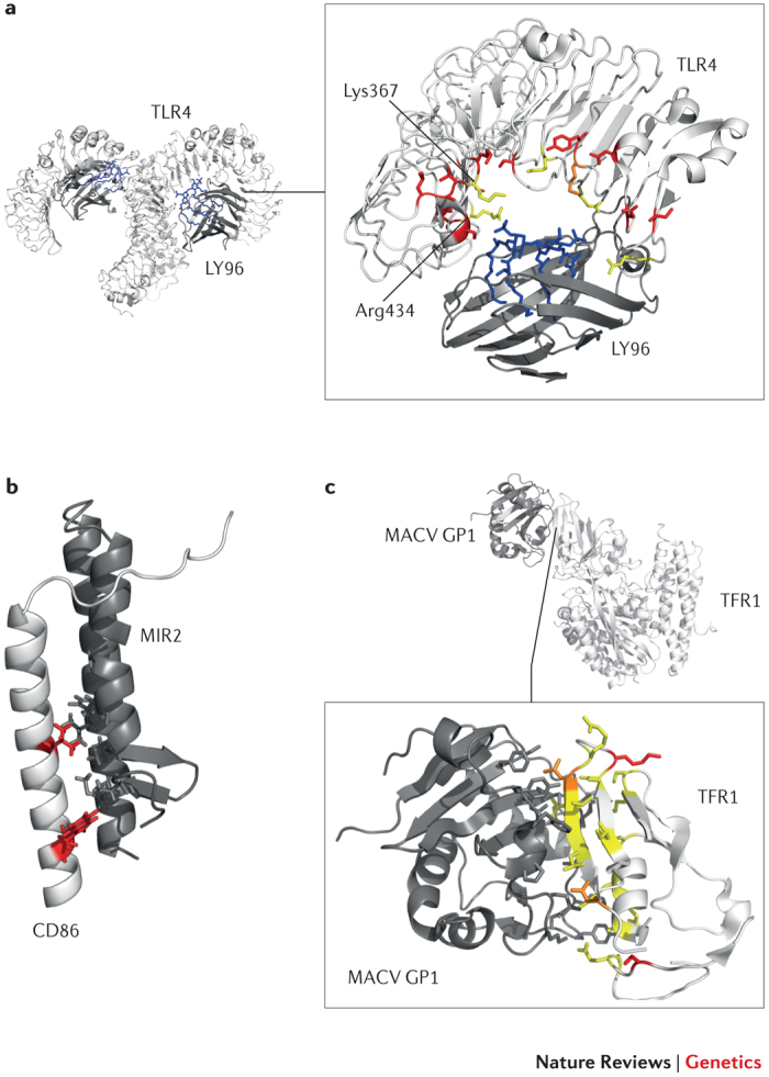
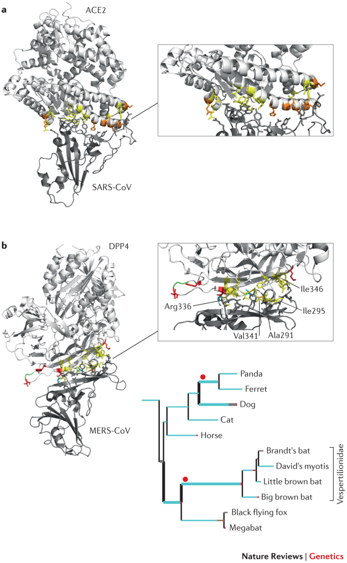
Infections are one of the major selective pressures acting on humans, and host-pathogen interactions contribute to shaping the genetic diversity of both organisms. Evolutionary genomic studies take advantage of experiments that natural selection has been performing over millennia. In particular, inter-species comparative genomic analyses can highlight the genetic determinants of infection susceptibility or severity. Recent examples show how evolution-guided approaches can provide new insights into host-pathogen interactions, ultimately clarifying the basis of host range and explaining the emergence of different diseases. We describe the latest developments in comparative immunology and evolutionary genetics, showing their relevance for understanding the molecular determinants of infection susceptibility in mammals.


Conflict of interest statement

The authors declare no competing financial interests.

Figures

Figure 1
Figure 1. Examples of positive selection at the host–pathogen interaction surface.
As discussed in Box 1, regions at the host–pathogen contact interface are expected to be targeted by the strongest selective pressure. Three examples are shown here. a | Detail of the Toll-like receptor 4 (TLR4)–lymphocyte antigen 96 (LY96)–lipid IVa complex. Mouse TLR4 and LY96 are in white and grey, respectively; lipid IVa is in blue. Sites that are positively selected in mammals are mapped onto the TLR4 structure (red): several of these flank or correspond (orange) to residues that differ between humans and mice and that surround the phosphate groups of lipid IVa (yellow). If Lys367 and Arg434 are replaced with the human residues (Glu369 and Gln436, respectively), the responsiveness of mouse TLR4–LY96 to lipid IVa is abolished. b | Structures of human CD86 (white; transmembrane and juxtamembrane region) and MIR2 (grey; encoded by Kaposi sarcoma-associated herpesvirus). CD86 sites that are involved in the interaction and that are positively selected in mammals are shown in red. c | Complex of transferrin receptor protein 1 (TFR1) with the surface glycoprotein (GP1) of Machupo virus (MACV), a rodent arenavirus that can also infect humans through zoonotic transmission. TFR1 residues involved in the interaction with GP1 are in yellow, positively selected sites are in red and positively selected sites that directly interact with GP1 are in orange. PowerPoint slide
Figure 2
Figure 2. Positive selection at the cellular receptors for coronaviruses (SARS-CoV and MERS-CoV).
The receptor-binding domains (RBDs) are structurally similar in the spike proteins of severe acute respiratory syndrome coronavirus (SARS-CoV) and Middle East respiratory syndrome coronavirus (MERS-CoV), but these proteins bind distinct cellular receptors. The structure of the SARS-CoV and MERS-CoV RBDs in complex with angiotensin-converting enzyme 2 (ACE2) and dipeptidyl peptidase 4 (DPP4), respectively, are shown with the binding interfaces enlarged. In both panels, sites that directly interact with the RBD are shown in yellow. a | ACE2 residues that are responsible for RBD binding and that are also positively selected in bats are shown in orange. b | DPP4 residues that are positively selected at the RBD binding interface are shown in red (positively selected) and orange (positively selected and interacting); sites in cyan were found to be positively selected along specific branches (Supplementary information S1,S2 (box, table)), as shown in the tree panel. The tree includes a subset of relevant branches, with those showing evidence of episodic positive selection represented with thick lines and red dots. Branch colours indicate the strength of selection (dN/dS): red indicates positive selection (dN/dS>5); blue indicates purifying selection (dN/dS=0); and grey indicates neutral evolution (dN/dS=1). Human residues that modify the binding energy if they are replaced with their hamster counterparts are labelled. One of these (Val341) is positively selected (orange). A three amino acid deletion in bats is shown in green (see Supplementary information S1,S2 (box, table)). PowerPoint slide
Figure 3
Figure 3. Genes involved in antigen processing and presentation and T cell regulation are common targets of positive selection in mammals.
All pathway components are designated using official gene names (excluding the major histocompatibility complex (MHC) and T cell receptor (TCR)) and are highlighted in red if they are targets of positive selection in mammals or primates,,. The molecular components of different antigen processing and presentation pathways are shown (details from Refs 107,108) to provide a general overview of the extent of positive selection and to highlight the function of positively selected genes, as most of their protein products directly interact with the antigen. Thus, the figure is not meant to show all molecules involved in the process or to convey mechanistic insights. Also, some genes may show tissue-specific expression or may be induced under specific circumstances: their products are nonetheless included for the sake of completeness. As for T cell regulatory molecules, the representation does not reflect the stoichiometry of binding (for example, CD28 functions as a dimer). Notably, the same molecule may be expressed by different populations of T cells, although here each molecule is shown on one T cell type only (to avoid redundancy). The dashed arrows and '?' indicate steps that lack clear molecular definition or are only inferred. The orange circles, and red and blue shapes at the bottom of the figure represent proteolytic fragments. B2M, β2-microglobulin; BLMH, bleomycin hydrolase; CALR, calreticulin; CD40LG, CD40 ligand; CTLA4, cytotoxic T lymphocyte protein 4; CTS, cathepsin; CYB, cytochrome b; ERAP, endoplasmic reticulum aminopeptidase; HAVCR2, hepatitis A virus cellular receptor 2; HLA-DM, major histocompatibility complex, class II, DM; ICOS, inducible T cell co-stimulator; ICOSLG, ICOS ligand; IFI30, interferon-γ-inducible protein 30; iNKT, invariant natural killer T; iTCR, invariant TCR; LGMN, legumain; LNPEP, leucyl-cystinyl aminopeptidase; NCF, neutrophil cytosol factor; NPEPPS, puromycin-sensitive aminopeptidase (also known as PSA); NRD1, nardilysin; PDCD1, programmed cell death 1; PDCD1LG2, programmed cell death 1 ligand 2; PDIA3, protein disulfide-isomerase A3; ROS, reactive oxygen species; TAP, antigen peptide transporter; TAPBP, TAP-binding protein (also known as tapasin); THOP1, thimet oligopeptidase 1; TPP2, tripeptidyl-peptidase 2. PowerPoint slide
Figure 4
Figure 4. Positive and purifying selection.
a | Distribution of dN/dS values for human–mouse one-to-one orthologues. The values for some of the genes discussed in this Review are indicated. Data were derived from the Ensembl BioMart database (see Further information). b | Natural selection acting on mammalian Niemann–Pick C1 (NPC1) genes. NPC1 is shown with its predicted membrane topology and protein regions coloured in hues of blue that represent the percentage of negatively selected sites (as detected by the single-likelihood ancestor counting method using Datamonkey); the darker the blue, the higher the percentage. The location of three positively selected residues (red) is indicated on the left, and an alignment of the corresponding region is shown on the protein to the right (with red and blue representing positively and negatively selected sites, respectively). The interaction with the glycoprotein (GP; green) of filoviruses (such as Ebola virus, Marburg virus or Lloviu virus) is shown. GP binds NPC1 after processing by cellular proteases. ACE2, angiotensin-converting enzyme 2; DARC, Duffy blood group, atypical chemokine receptor; MX1, myxovirus resistance 1; SSD, sterol-sensing domain; TFRC, transferrin receptor; TLR4, Toll-like receptor 4. PowerPoint slide

Similar articles

Cited by

References

    1. Fumagalli M, et al. Signatures of environmental genetic adaptation pinpoint pathogens as the main selective pressure through human evolution. PLoS Genet. 2011;7:e1002355. - PMC - PubMed
    1. Van Valen L. A new evolutionary law. Evol. Theory. 1973;1:1–30.
    1. Brockhurst, M. A. et al. Running with the Red Queen: the role of biotic conflicts in evolution. Proc. Biol. Sci.10.1098/rspb.2014.1382 (2014). - PMC - PubMed
    1. Haldane JBS. The Causes of Evolution. 1932.
    1. Samson JE, Magadan AH, Sabri M, Moineau S. Revenge of the phages: defeating bacterial defences. Nature Rev. Microbiol. 2013;11:675–687. - PubMed

Publication types

LinkOut - more resources