. 2015 Apr;56(4):2486-97.
doi: 10.1167/iovs.15-16509.

Ornithine-δ-Aminotransferase Inhibits Neurogenesis During Xenopus Embryonic Development

Affiliations

Ornithine-δ-Aminotransferase Inhibits Neurogenesis During Xenopus Embryonic Development

Ying Peng et al. Invest Ophthalmol Vis Sci. 2015 Apr.

Abstract

Purpose: In humans, deficiency of ornithine-δ-aminotransferase (OAT) results in progressive degeneration of the neural retina (gyrate atrophy) with blindness in the fourth decade. In this study, we used the Xenopus embryonic developmental model to study functions of the OAT gene on embryonic development.

Methods: We cloned and sequenced full-length OAT cDNA from Xenopus oocytes (X-OAT) and determined X-OAT expression in various developmental stages of Xenopus embryos and in a variety of adult tissues. The phenotype, gene expression of neural developmental markers, and enzymatic activity were detected by gain-of-function and loss-of-function manipulations.

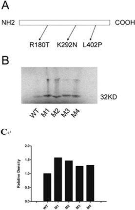
Results: We showed that X-OAT is essential for Xenopus embryonic development, and overexpression of X-OAT produces a ventralized phenotype characterized by a small head, lack of axial structure, and defective expression of neural developmental markers. Using X-OAT mutants based on mutations identified in humans, we found that substitution of both Arg 180 and Leu 402 abrogated both X-OAT enzymatic activity and ability to modulate the developmental phenotype. Neurogenesis is inhibited by X-OAT during Xenopus embryonic development.

Conclusions: Neurogenesis is inhibited by X-OAT during Xenopus embryonic development, but it is essential for Xenopus embryonic development. The Arg 180 and Leu 402 are crucial for these effects of the OAT molecule in development.


Figures

Figure 1
Figure 1
Comparison of human and Xenopus OAT amino acid sequences. Arrows indicate locations of X-OAT mutations generated as described in the Methods section.
Figure 2
Figure 2
Expression of X-OAT at different developmental stages of Xenopus and in different tissues of adult Xenopus. Reverse transcriptase PCR analysis of X-OAT expression: (A) brain, lane 1; liver, lane 2; kidney, lane 3; heart, lane 4; testis, lane 5; lung, lane 6; muscle, lane 7; stomach, lane 8; small intestine, lane 9; large intestine, lane 10; dorsal skin, lane 11; ventral skin, lane 12. No RTs (lane 13) were performed with total RNAs obtained from the brain tissue and processed through the reactions without RT to confirm the absence of contaminating genomic DNA. Expression of EF-1α was tested to serve as a loading control. (B) Oocyte (stages 5–6), lane 1; stage 4, lane 2; stage 6, lane 3; stage 9, lane 4; stage 10, lane 5; stage 11, lane 6; stage 13, lane 7; stage 15, lane 8; stage 17, lane 9; stage 25, lane 10; stage 30, lane 11; stage 40, lane 12. No RT control reactions (lane 13) were performed with total RNAs obtained from the oocyte.
Figure 3
Figure 3
Spatio-temporal expression of OAT during embryonic development. Whole-mount in situ hybridization analysis shows that (A) X-OAT is expressed in all regions of oocyte. (B) In the early neural stage, X-OAT is localized principally to the dorsal side, especially in the notoplate (white arrow). (C) The X-OAT is predominantly expressed in brain and spinal cord at tailbud stage, as shown with digoxigenin-labeling (white arrow).
Figure 4
Figure 4
Deletion of endogenous X-OAT produces gastrulation defect and death of embryo. Either 5 ng or 10 ng of X-OAT-Mo was injected into the animal pole area of two-cell stage embryos. Embryos were cultured in 30% MMR solution and photographed at the equivalent of stage 13: (A) normal control; (B) injected with 5 ng X-OAT-Mo; (C) injected with 10 ng X-OAT-Mo photographed at stage 10.
Figure 5
Figure 5
Overexpression of X-OAT-induced morphological change of Xenopus embryo. (A) Top: mRNA encoding β-gal (1 ng) served as negative control; bottom: X-OAT sense RNA (1 ng) was injected into animal pole area at the two-cell stage. (B) Top: mRNA encoding β-gal (1 ng) served as negative control; bottom: X-OAT sense RNA (1 ng) was injected into dorsal marginal zone of four-cell stage embryos.
Figure 6
Figure 6
The OAT inhibited neuralization induced by activin and retinoic acid in Xenopus ACs. Two-cell stage embryos were injected in the animal pole area with mRNA (1 ng) encoding (A) β-gal, (B) sense X-OAT, or (C) X-OAT-Mo. The ACs were dissected at stages 8.5 to 9.0 and cultured in 67% Lebovitz's L-15 medium with activin (10 ng/mL) and retinoic acid (10−5 M) until they reached the equivalent of stage 22; three ACs of each group for photography. (A) All eight ACs appear slightly swollen with one side white and the other black (100%); (B) seven ACs appear mostly round and evenly brownish (88%), one AC has died; (C) the phenotype of all eight ACs similar to the (A) group's phenotype (100%).
Figure 7
Figure 7
Inhibition of NCAM, HoxB9, and Otx2 expression by X-OAT in ACs cultured with activin and retinoic acid. The animal pole area of two-cell stage embryos was injected with mRNA encoding β-gal (1 ng), X-OAT, sense (s) (1 ng); X-OAT, sense (s) (1 ng) + X-OAT-Mo (as) (5 ng). The ACs were dissected at stages 8.5 to 9.0 and cultured in 67% Leibovitz's L-15 medium containing activin (10 ng/mL) and retinoic acid (10−5 M) until equivalent of stage 22. Total RNA was isolated from the ACs and assayed for expression by RT-PCR (A) for expression of NCAM, and (B) for expression of Otx2 and HoxB9. Expression of EF-1α was used as loading control. Total RNAs from a whole embryo at the equivalent stage were used as a positive control. Controls with “No RT” reactions were performed with total RNAs obtained from the embryo and processed through the reactions without RT to confirm the absence of contaminating genomic DNA.
Figure 8
Figure 8
Translation of WT and mutated X-OATs. The introduced mutations are shown with their designations (A). The M2 has both the M1 and M3 mutations. M1: X-OATR180T; M2: X-OATR180T/L402P; M3: X-OATL402P; M4: X-OATK292N; WT: X-OATWT. Synthetic WT and mutated X-OAT mRNA were translated in a TNT-coupled reticulocyte lysate system (Promega) with [35S] methionine. Products were analyzed by autoradiography on an SDS Tris-HCl gel. A 32-kDa band appeared at similar intensity in all the lanes loaded with the translation products (B) and the densitometric analysis (C).
Figure 9
Figure 9
Phenotype observations and RT-PCR analysis of ACs expressing WT and mutated X-OAT; β-gal or 1 ng mRNA encoding WT or each of the X-OAT mutants was injected into the animal pole at the two-cell stage. (A) Embryos were grown to tadpole stage for photography. (a) β-gal (normal: 48/50, died: 2/50), (b) X-OATWT (normal: 8/50, ventralization: 34/50, died: 8/50), (c) X-OATR180T (M1) (normal: 8/50, ventralization: 37/50, died: 5/50), (d): X-OATR 180T/L402P (M2) (normal: 46/50, died: 4/50), (e) X-OATL402P (M3) (normal: 10/50, ventralization: 31/50, died: 9/50), or (f) X-OATK292N (M4) (normal: 7/50, ventralization: 36/50, died: 7/50). (B) The ACs were dissected at stage 8.5 to 9.0 and cultured to equivalent of stage 22. The ACs were harvested for photography: (a) β-gal (neurolization 8/8), (b) X-OATWT (normal: 8/8), (c) X-OATR180T (M1) (normal: 8/8), (d) X-OATR 180T/L402P (M2) (neurolization: 7/8, died: 1/8), (e) X-OATL402P (M3) (normal: 8/8) or (f) X-OATK292N (M4) (normal: 7/8, died: 1/8), or (C) for RT-PCR analysis of NCAM expression with EF-1α as control.
Figure 10
Figure 10
Differential X-OAT enzyme activities in ACs expressing β-gal, X-OAT-Mo, X-OATWT, and mutants. Messenger RNA (1 ng) from X-OATWT or mutants was injected into the animal pole at the two-cell stage. The ACs were dissected at stage 8.5 to 9.0 and cultured to equivalent of stage 22. The ACs were harvested in PBS buffer (16 ACs per group) for protein concentration measurement and X-OAT enzyme activity assay (see Methods). All groups were assayed in triplicate. P values are compared between β-gal and each OAT by Student's t-tests. *P < 0.01, compared with β-gal; **P < 0.001, compared with β-gal.

Similar articles

Cited by

References

    1. Hagedorn CH,, Phang JM. Catalytic transfer of hydride ions from NADPH to oxygen by the interconversions of proline and delta 1-pyrroline-5-carboxylate. Arch Biochem Biophys. 1986; 248: 166–174. - PubMed
    1. Phang JM. The regulatory functions of proline and pyrroline-5-carboxylic acid. Curr Top Cell Regul. 1985; 25: 91–132. - PubMed
    1. Fremeau RT, Jr,, Caron MG,, Blakely RD. Molecular cloning and expression of a high affinity L-proline transporter expressed in putative glutamatergic pathways of rat brain. Neuron. 1992; 8: 915–926. - PubMed
    1. Renick SE,, Kleven DT,, Chan J,, et al. The mammalian brain high-affinity L-proline transporter is enriched preferentially in synaptic vesicles in a subpopulation of excitatory nerve terminals in rat forebrain. J Neurosci. 1999; 19: 21–33. - PMC - PubMed
    1. Gogos JA,, Santha M,, Takacs Z,, et al. The gene encoding proline dehydrogenase modulates sensorimotor gating in mice. Nat Genet. 1999; 21: 434–439. - PubMed

Publication types

MeSH terms

LinkOut - more resources