

Viral Vector Effects on Exoenzyme C3 Transferase-Mediated Actin Disruption and on Outflow Facility
- PMID: 25783606
- PMCID: PMC4408885
- DOI: 10.1167/iovs.14-15909
Viral Vector Effects on Exoenzyme C3 Transferase-Mediated Actin Disruption and on Outflow Facility
Abstract
Purpose: Purified Clostridium botulinum exoenzyme C3 transferase (C3) effects on the actin cytoskeleton in human trabecular meshwork cells (HTM) and on the outflow facility response in monkey organ-cultured anterior segments (MOCAS) were determined in the presence or absence of viral vectors.
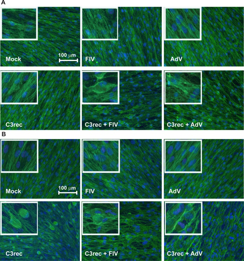
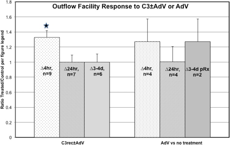
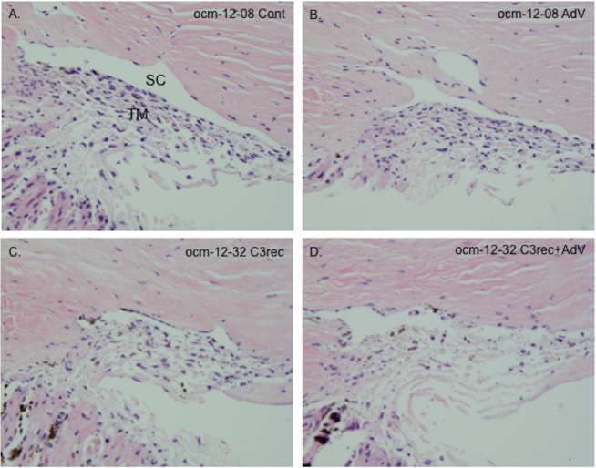
Methods: Human adenovirus type 5 (AdV) and feline immunodeficiency virus (FIV) vectors were produced using kits. Cell soluble purified C3 (C3cs) was purchased commercially. Recombinant C3 (C3rec) cDNA was overexpressed in Escherichia coli and purified. The HTM cells were incubated with up to 10 μg/mL C3cs or with 5 μg of C3rec and/or viral vector (multiplicity of infection [MOI] = 25). Cells then were fixed and stained for actin. Outflow facility in MOCAS was measured at baseline, 4 hours, 24 hours, and 3 to 4 days following bolus injection of AdV (1.6 × 10⁷ transducing units) and/or 2.5 μg C3rec.
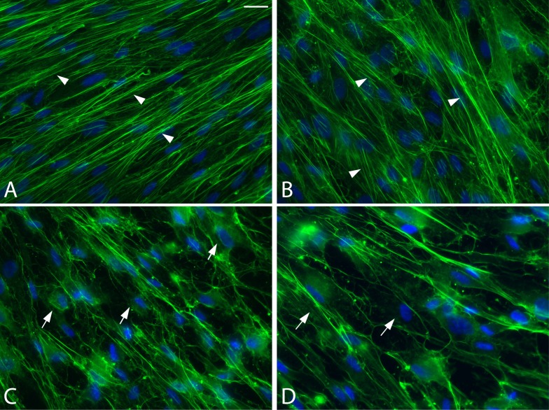
Results: The HTM cells treated for 4 hours with C3cs (all doses) or for 24 hours with C3rec developed a rounded morphology and lost stress fibers. Cells transduced with vectors alone showed no changes at any time point. Cells exposed to C3rec and cotransduced with either viral vector showed significant disruption of the actin cytoskeleton within 4 hours after exposure, which persisted at 24 hours. In MOCAS, the AdV vector alone had no effect on outflow facility, but enhanced the response to C3rec at 4 hours.
Conclusions: Coadministration of viral vectors enhances the ability of C3 transferase to disrupt actin stress fiber formation in HTM cells and increase outflow facility in MOCAS. Viral vectors potentially could be used to increase the bioavailability of proteins for cells that are difficult to transfect.
Figures
Similar articles
-
The effect of C3 transgene expression on actin and cellular adhesions in cultured human trabecular meshwork cells and on outflow facility in organ cultured monkey eyes.Mol Vis. 2005 Dec 13;11:1112-21. Mol Vis. 2005. PMID: 16379023
-
Preservation of aqueous outflow facility after second-generation FIV vector-mediated expression of marker genes in anterior segments of human eyes.Invest Ophthalmol Vis Sci. 2002 Dec;43(12):3686-90. Invest Ophthalmol Vis Sci. 2002. PMID: 12454037
-
Caldesmon transgene expression disrupts focal adhesions in HTM cells and increases outflow facility in organ-cultured human and monkey anterior segments.Exp Eye Res. 2006 Jun;82(6):935-44. doi: 10.1016/j.exer.2005.12.002. Epub 2006 Jan 26. Exp Eye Res. 2006. PMID: 16442523
-
COCH transgene expression in cultured human trabecular meshwork cells and its effect on outflow facility in monkey organ cultured anterior segments.Invest Ophthalmol Vis Sci. 2010 Apr;51(4):2060-6. doi: 10.1167/iovs.09-4521. Epub 2009 Nov 20. Invest Ophthalmol Vis Sci. 2010. PMID: 19933177 Free PMC article.
-
Gene transfer of dominant-negative RhoA increases outflow facility in perfused human anterior segment cultures.Mol Vis. 2002 Mar 6;8:32-44. Mol Vis. 2002. PMID: 11889464
Cited by
-
Lentiviral Vector-Mediated Expression of Exoenzyme C3 Transferase Lowers Intraocular Pressure in Monkeys.Mol Ther. 2019 Jul 3;27(7):1327-1338. doi: 10.1016/j.ymthe.2019.04.021. Epub 2019 May 9. Mol Ther. 2019. PMID: 31129118 Free PMC article.
-
Knockdown of TRIM5α or TRIM11 increases lentiviral vector transduction efficiency of human Muller cells.Exp Eye Res. 2021 Mar;204:108436. doi: 10.1016/j.exer.2021.108436. Epub 2021 Jan 10. Exp Eye Res. 2021. PMID: 33440192 Free PMC article.
-
Deconstructing aqueous humor outflow - The last 50 years.Exp Eye Res. 2020 Aug;197:108105. doi: 10.1016/j.exer.2020.108105. Epub 2020 Jun 23. Exp Eye Res. 2020. PMID: 32590004 Free PMC article. Review.
-
Role of the Rho GTPase/Rho kinase signaling pathway in pathogenesis and treatment of glaucoma: Bench to bedside research.Exp Eye Res. 2017 May;158:23-32. doi: 10.1016/j.exer.2016.08.023. Epub 2016 Sep 1. Exp Eye Res. 2017. PMID: 27593914 Free PMC article. Review.
-
Lentiviral Vectors for Ocular Gene Therapy.Pharmaceutics. 2022 Jul 31;14(8):1605. doi: 10.3390/pharmaceutics14081605. Pharmaceutics. 2022. PMID: 36015231 Free PMC article. Review.
References
-
- Llobet A,, Gasull X,, Gual A.Understanding trabecular meshwork physiology: a key to the control of intraocular pressure? News Physiol Sci. 2003; 18: 205–209. - PubMed
-
- Hall A.Rho GTPases and the actin cytoskeleton. Science. 1998; 279: 509–514. - PubMed
-
- Aktories K,, Mohr C,, Koch G.Clostridium botulinum C3 ADP-ribosyltransferase. Curr Top Microbiol Immunol. 1992; 175: 115–131. - PubMed
-
- Liu X,, Hu Y,, Filla MS,, et al. The effect of C3 transgene expression on actin and cellular adhesions in cultured human trabecular meshwork cells and on outflow facility in organ cultured monkey eyes. Mol Vis. 2005; 11: 1112–1121. - PubMed
Publication types
MeSH terms
Substances
Grants and funding
LinkOut - more resources
Full Text Sources
Other Literature Sources
Miscellaneous
