. 2015 Nov;8(6):1262-74.
doi: 10.1038/mi.2015.16. Epub 2015 Mar 18.

Inhibition of CD23-mediated IgE transcytosis suppresses the initiation and development of allergic airway inflammation

Affiliations

Inhibition of CD23-mediated IgE transcytosis suppresses the initiation and development of allergic airway inflammation

S Palaniyandi et al. Mucosal Immunol. 2015 Nov.

Abstract

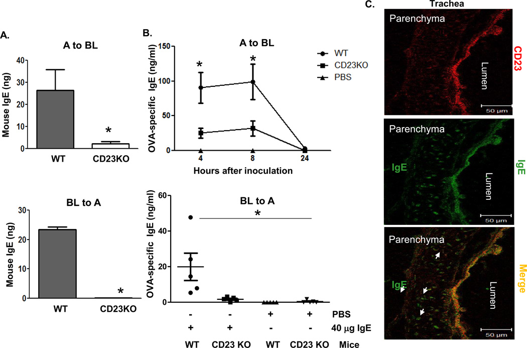
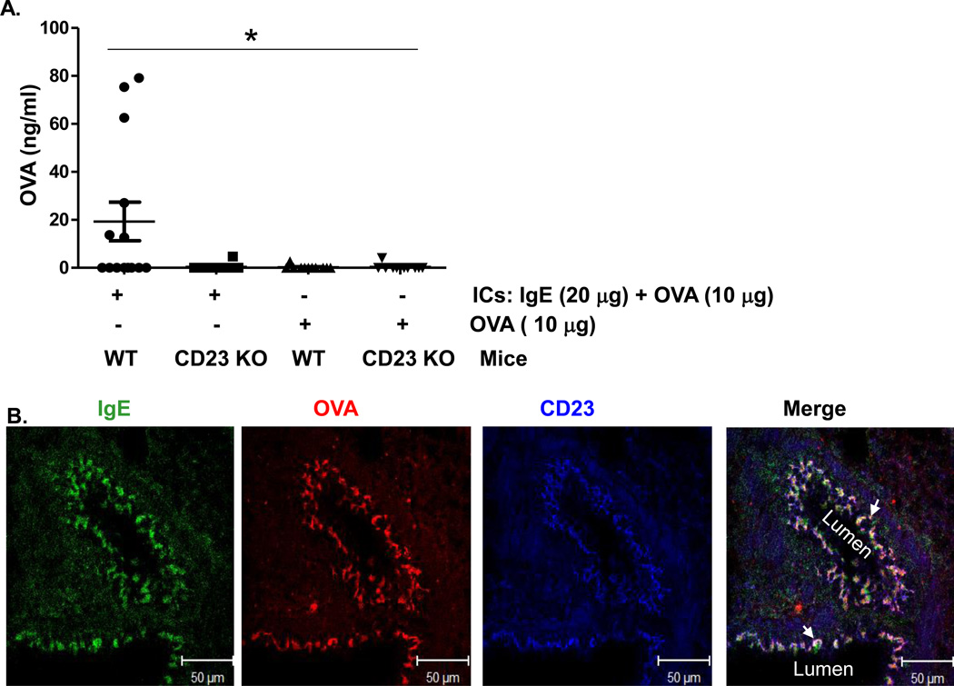
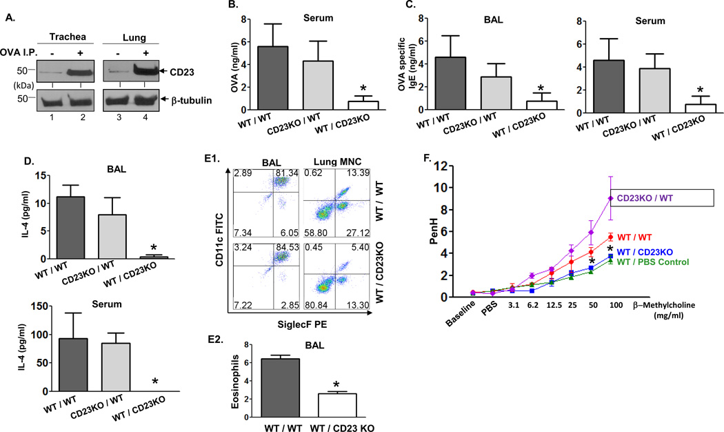
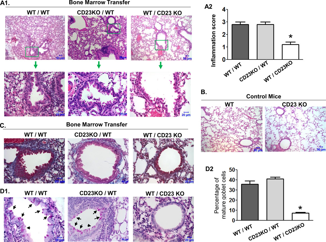
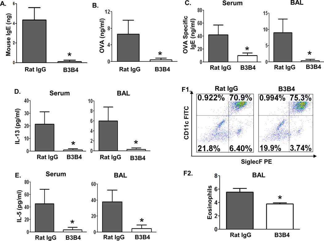
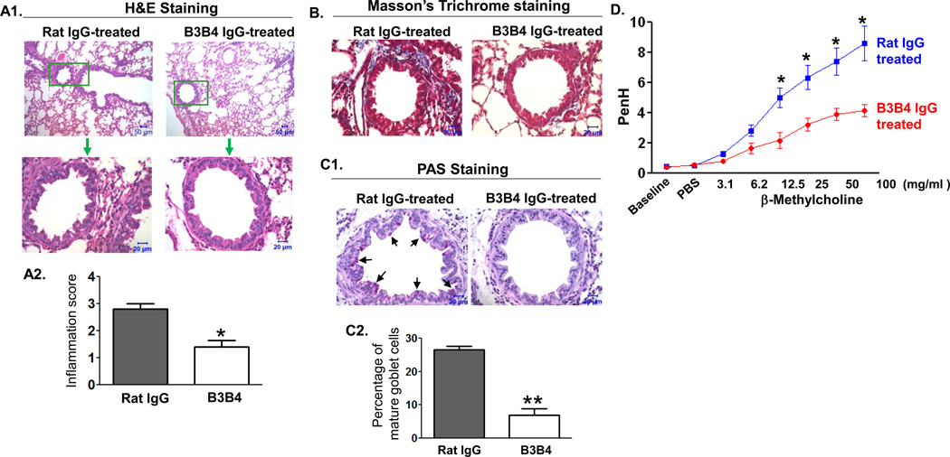
The epithelial lining of the airway tract and allergen-specific IgE are considered essential controllers of inflammatory responses to allergens. The human low affinity IgE receptor, CD23 (FcɛRII), is capable of transporting IgE or IgE-allergen complexes across the polarized human airway epithelial cell (AEC) monolayer in vitro. However, it remains unknown whether the CD23-dependent IgE transfer pathway in AECs initiates and facilitates allergic inflammation in vivo, and whether inhibition of this pathway attenuates allergic inflammation. To this end, we show that in wild-type (WT) mice, epithelial CD23 transcytosed both IgE and ovalbumin (OVA)-IgE complexes across the airway epithelial barrier, whereas neither type of transcytosis was observed in CD23 knockout (KO) mice. In chimeric mice, OVA sensitization and aerosol challenge of WT/WT (bone-marrow transfer from the WT to WT) or CD23KO/WT (CD23KO to WT) chimeric mice, which express CD23 on radioresistant airway structural cells (mainly epithelial cells) resulted in airway eosinophilia, including collagen deposition and a significant increase in goblet cells, and increased airway hyperreactivity. In contrast, the absence of CD23 expression on airway structural or epithelial cells, but not on hematopoietic cells, in WT/CD23KO (the WT to CD23KO) chimeric mice significantly reduced OVA-driven allergic airway inflammation. In addition, inhalation of the CD23-blocking B3B4 antibody in sensitized WT mice before or during airway challenge suppressed the salient features of asthma, including bronchial hyperreactivity. Taken together, these results identify a previously unproven mechanism in which epithelial CD23 plays a central role in the development of allergic inflammation. Further, our study suggests that functional inhibition of CD23 in the airway is a potential therapeutic approach to inhibit the development of asthma.


Figures

Fig 1
Fig 1. Expression of CD23 in mouse AECs
Cell lysates (50 µg) derived from primary nasal (A), tracheal (B), and lung (B) epithelial cells were electrophoresed on a 12 % SDS-PAGE gel under reducing condition. The separated proteins were transferred onto a nitrocellulose membrane, blotted with rat anti-mouse CD23 mAb (B3B4) followed by HRP-conjugated rabbit anti-rat IgG Ab. Proteins were visualized using the ECL method. Lysates (50 µg) from mouse spleen or CHO cells were used as a positive or negative control, respectively. β-tubulin was used as an internal control. Arrows indicate mouse CD23 and β-tubulin. (C) Immunohistochemical staining of mouse lung. Normal mouse lung was prepared in OCT medium and cryosectioned at 5 µm. Frozen tissue sections were fixed and permeabilized with ice-cold acetone and blocked with 10% normal goat serum. A spleen section was used as a positive control. Sections were incubated with rabbit anti-CD23 Ab or normal rabbit IgG, followed by staining with Alexa Fluor 555-conjugated goat anti-rabbit Ab. Nuclei were stained with DAPI. Images were captured using a Zeiss LSM510 confocal microscope. Samples were visualized under consisten contrast and brightness settings.
Fig 2
Fig 2. CD23-mediated transcytosis of IgE across mouse epithelial monolayers
A. Transcytosis of mouse IgE in primary mouse tracheal epithelial cells (TEC). TEC isolated from WT or CD23 KO mice were grown on transwell filters and cells were allowed to become polarized. Mouse IgE was added to the apical (top panel) or basolateral (lower panel) chambers and incubated at 37°C for 2 h. The medium from opposite chamber was collected and IgE concentration was measured by ELISA. A: Apical; BL: Basolateral. *P<0.05. B. Airway transcytosis of mouse IgE in wild type (WT) or CD23 KO mice. Top Panel: OVA-specific IgE (40 µg) was i.n. inoculated into either WT or CD23 KO mice for 5 min. Sterile PBS was used as a control. Sera were collected at indicated time points. Bottom Panel: OVA-specific IgE (40 µg) or sterile PBS was i.p. inoculated into either WT or CD23 KO mice. BAL fluid was collected 8 h after injection. OVA-specific IgE was measured in the sera and BAL fluid by ELISA. *P<0.05. C. Colocalization of CD23 and IgE in mouse trachea. Naive mice were anaesthetized with avertin and 20 µg of mouse IgE or PBS was i.n. inoculated. Mice were sacrificed 20 min after treatment, and the trachea was collected and snap frozen in OCT medium and cryosectioned at 5 µm. Frozen tissue sections were fixed and permeabilized with ice-cold acetone and blocked with 10% normal goat serum (NGS). Sections were incubated with rabbit anti-CD23 Ab followed by staining with Alexa Fluor 555-conjugated goat anti-rabbit Ab (red) and FITC-conjugated goat anti-mouse IgE Ab (green). Lung epithelial cells did not stain positive with rabbit anti-IgE Ab. Images were captured using a LSM510 confocal microscope. Samples were visualized under consistent contrast and brightness settings. Arrows indicate IgE Ab transported into the parenchyma.
Fig 3
Fig 3. CD23-mediated transcytosis of OVA-IgE immune complexes (ICs) across mouse epithelial monolayers
(A). Apical to basolateral transcytosis of ICs in naive mice. ICs were formed with 20 µg OVA-specific IgE and 10 µg of OVA at room temperature for 30 min. WT or CD23 KO mice were anaesthetized with avertin and ICs, 10µg OVA, or PBS was i.n. inoculated, respectively. Sera were collected 8 h later and OVA antigen quantitated by ELISA. *P<0.05. (B). Colocalization of CD23 and ICs in the lungs of naïve mice. OVA-IgE ICs or PBS were i.n. inoculated into anaesthetized mice, which were sacrificed 20 min later and lung tissue was prepared in OCT medium and cryosectioned at 5 µm. Frozen sections were fixed and permeabilized with ice-cold acetone and blocked with 10% NGS. Sections were incubated with rabbit anti-CD23 Ab or mouse anti-chicken OVA Ab followed by staining with Alexa fluor 633-conjugated goat anti-rabbit Ab, Alexa fluor 555-conjugated goat anti-mouse Ab, and FITC conjugated goat anti-mouse IgE Ab. Nuclei were stained with DAPI and sections were imaged using a LSM510 confocal microscope. Samples were visualized under consistent contrast and brightness settings. Arrows indicate the colocalization (white).
Fig 4
Fig 4. Epithelial CD23 facilitates airway inflammation after OVA sensitization and challenge in chimeric mice
A. Mice were sensitized (i.p.) with OVA or left untreated. Lung and trachea epithelial cells were isolated and cell lysates (50 µg) were subjected to 12 % SDS-PAGE gel electrophoresis under reducing conditions. Proteins were transferred onto nitrocellulose membranes and blotted with rat-anti mouse CD23 mAb (B3B4) followed by HRP-conjugated rabbit anti-rat IgG Ab. Proteins were visualized using ECL. Arrows indicate mouse CD23 and β-tubulin. B–D. WT/WT, CD23KO/WT, WT/CD23KO chimeric mice were i.p. sensitized with 100 µg OVA plus 4 mg alum at day 0 and subsequently injected (i.p.) with 100 µg OVA on day 7 and 14. Mice were challenged on day 21 with nebulized 1 % OVA for 30 min. 5 h after OVA challenge, sera were collected and OVA quantitated by ELISA (B). 24 h after OVA challenge, BAL fluid and sera were collected and OVA-specific IgE (C) and IL-4 (D) concentrations assessed by ELISA. *P<0.05. E. Flow cytometric analysis of eosinophils in BAL fluid. Chimeric mice were sensitized with OVA and received a single aerosol OVA challenge. Total leukocytes obtained from BAL fluid were gated on CD45+ CD11bhi/int and mononuclear cells obtained from lung were gated on CD45+ CD11bhi cells and further analyzed for CD11c and Siglec-F expression (E1). The mean percentage of eosinophils in BAL fluid from three different experiments was calculated (E2). *P<0.05. F. Airway responsiveness to methacholine (MCh) in chimeric and control mice. WT/WT, CD23KO/WT, and WT/CD23KO chimeric mice were sensitized (i.p.) with OVA and received a single aerosol challenge with OVA while control mice were sensitized and received a single aerosol challenge with PBS. Airway resistance was measured 24 h after challenge. First, the baseline and resistance against PBS challenge were measured followed by generation of a dose-response curve against an increasing concentration of nebulized MCh (3–100 mg/ml). *P<0.05.
Fig 5
Fig 5. Effects of epithelial CD23 on pathological changes in lung
WT/WT, CD23KO/WT, and WT/CD23KO chimeric mice were sensitized with OVA and received a single aerosol OVA challenge. Lung tissues were fixed in formalin and embedded in paraffin 24 h after OVA challenge. A. Lung tissue section were stained with H&E and examined by light microscopy at the indicated magnifications. Lung sections from WT/CD23KO chimeric mice (top right) showed a significant attenuation of inflammation compared with that from WT/WT (top left) and CD23KO/WT (top middle) mice. Insets (top, green squares) are shown at a higher amplification (arrow). Pronounced alveolar and bronchiolar changes are shown in WT/WT or CD23KO/WT mice. Semi-quantitation of inflammation by histological analysis (A2). *P<0.05. Lung sections from either CD23 KO or WT mice are shown in panel (B). C. Lung sections were stained using the Masson's trichrome method. WT/CD23KO chimeric mice showed significantly decreased peribronchial fibrosis (blue) in the lung compared with WT/WT or CD23KO/WT chimeric mice. Data are representative of two independent experiments. D. Lung sections were stained with PAS. WT/CD23 KO chimeric mice showed a significant reduction in goblet-cell hyperplasia (arrows) in the lung bronchial area (top panel) in comparison with that of WT/WT or CD23KO/WT chimeric mice (D1). Goblet cells were counted and the mean percentage of three different sections is indicated (D2). *P<0.05.
Fig 6
Fig 6. Effect of B3B4 Ab targeting of airway CD23 on IgE and allergen transcytosis, inflammatory cytokine production, and eosinophil infiltration
A. B3B4 Ab blocked IgE transcytosis in primary mouse TEC. Cells were isolated from WT mice, grown on transwell filters, and allowed to polarized. Purified B3B4 Ab or rat IgG2a (50 µg/ml) was added into the apical chamber for 45 min at 4°C to allow Ab binding to apical CD23. Mouse IgE was then added to apical chambers and incubated at 37°C for 2 h. Medium from the basolateral chamber was collected and IgE content measured by ELISA. *P<0.05. B–F. Sensitization of mice with OVA. Before challenge, OVA-sensitized WT mice received i.n. inoculation with 75 µg B3B4 Ab or rat IgG2a in PBS twice, once at 24 h before, and again 1 h before, challenge. Sera were collected 5 h after the single OVA challenge and OVA in sera was quantified by ELISA (B). BAL fluid or sera were collected 48 h after OVA challenge. Concentrations of OVA-specific IgE (C), IL-13 (D), and IL-5 (E) in either BAL fluid or sera were measured by ELISA. Eosinophils were assessed by flow cytometric analysis of CD45+ CD11bhi/int CD11c Siglec-F+ cells in BAL fluid. The mean percentage of the eosinophils in the BAL fluid from three different experiments was calculated (F2). *P<0.05.
Fig 7
Fig 7. Effect of B3B4 Ab targeting of airway CD23 on airway allergic inflammation
WT mice were sensitized with OVA and received i.n. inoculation of 75 µg B3B4 Ab or rat IgG2a in PBS twice, once at 24 h before and again 1 h before challenge. Subsequently, mice were challenged with OVA and 24 h later AHR was measured, and 48 h later mice were sacrificed and lung tissues were fixed in formalin and embedded in paraffin. A. Lung sections were stained with H&E and examined by light microscopy at the indicated magnifications. Lung sections in B3B4 Ab-treated mice showed significant attenuation of inflammation in comparison with that of control rat IgG2a-treated mice. Insets (green squares) are shown at higher amplifications. Semi-quantitative assessment of inflammation in histological sections was analyzed (A2 panel). *P<0.05. B. Lung tissue sections were stained using the Masson's trichrome method. B3B4 Ab-treated mice showed significantly decreased peribronchial fibrosis (blue) in the lung compared with control Ab-treated mice. C. Lung tissue sections were stained with PAS. B3B4-treated mice had a significant reduction in goblet-cell hyperplasia in the lung bronchial area compared with that of the rat IgG2a-treated mice (c1). Goblet cells were counted and the mean percentage of three different sections is presented (c2). *P<0.01. D. Airway responsiveness to methacholine (MCh) in B3B4 Ab-treated mice. AHR was measured 24 h after a single OVA challenge. First, baseline airway resistance and resistance against PBS challenge were measured and then a dose response curve against increasing concentrations of nebulized MCh (3–100 mg/ml) was generated. *P<0.05.

Similar articles

Cited by

References

    1. Galli SJ, Tsai M, Piliponsky AM. The development of allergic inflammation. Nature. 2008;454:445–454. - PMC - PubMed
    1. Licona-Limón P, Kim LK, Palm NW, Flavell RA. TH2, allergy and group 2 innate lymphoid cells. Nat. Immunol. 2013;14:536–542. - PubMed
    1. Crimi E, Scordamaglia A, Crimi P, Zupo S, Barocci S. Total and specific IgE in serum, bronchial lavage and bronchoalveolar lavage of asthmatic patients. Allergy. 1983;38:553–559. - PubMed
    1. Balzar S, Strand M, Rhodes D, Wenzel SE. Local IgE expression pattern in lung: relation to systemic IgE and asthma phenotypes. J. Allergy Clin. Immunol. 2007;119:855–862. - PMC - PubMed
    1. Fiset PO, Cameron L, Hamid Q. Local isotype switching to IgE in airway mucosa. J Allergy Clin Immunol. 2005;116:233–236. - PubMed

Publication types

MeSH terms