. 2015 Jul:325:12-26.
doi: 10.1016/j.heares.2015.03.002. Epub 2015 Mar 14.

Aminoglycoside ototoxicity and hair cell ablation in the adult gerbil: A simple model to study hair cell loss and regeneration

Affiliations

Aminoglycoside ototoxicity and hair cell ablation in the adult gerbil: A simple model to study hair cell loss and regeneration

Leila Abbas et al. Hear Res. 2015 Jul.

Abstract

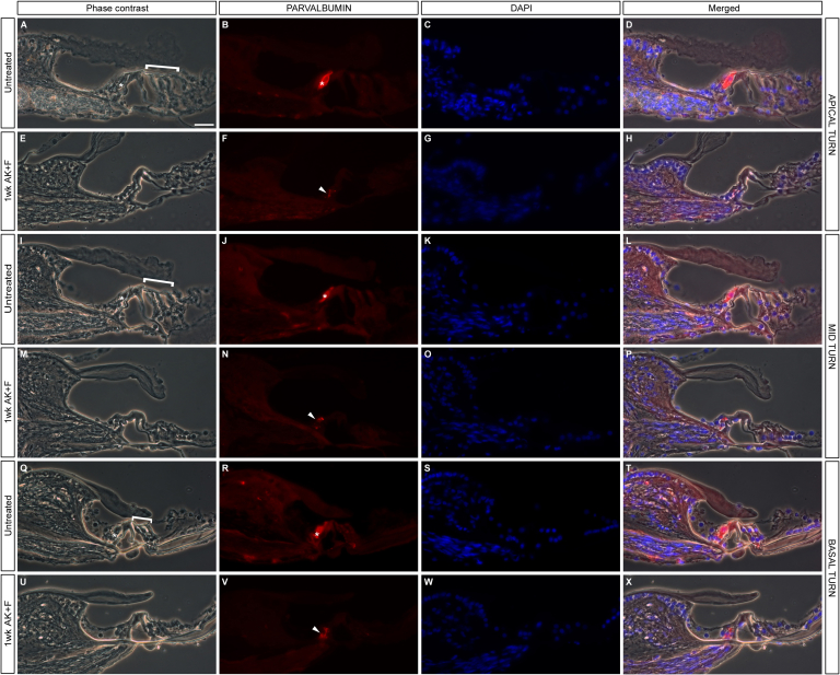
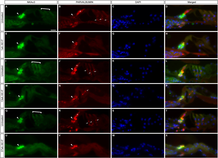
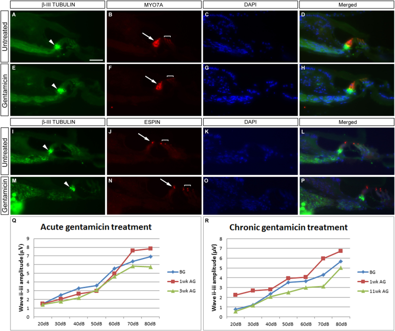
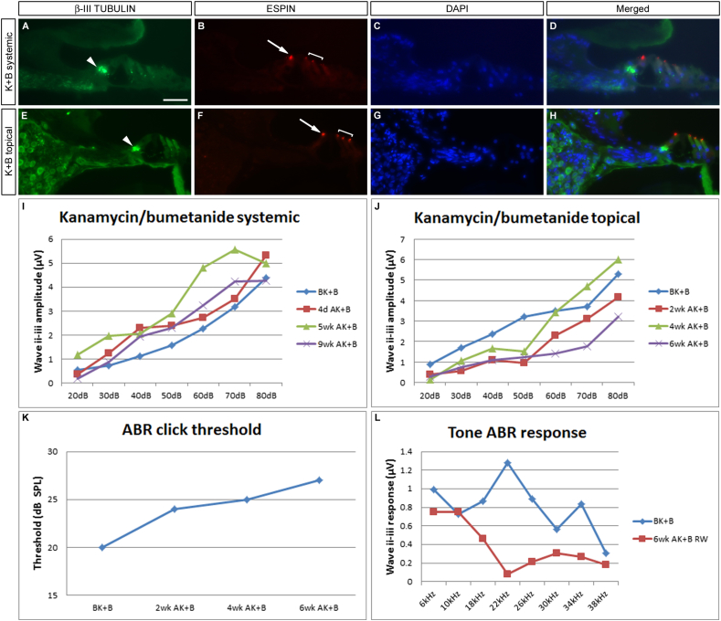
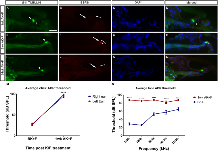
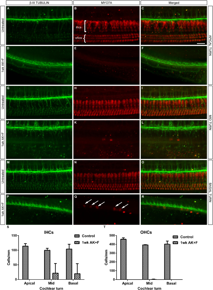
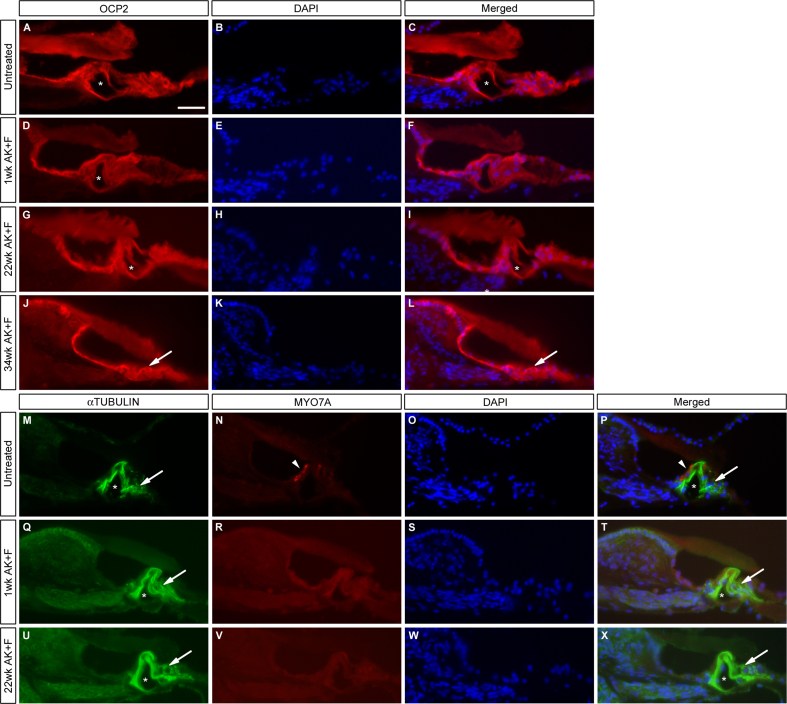
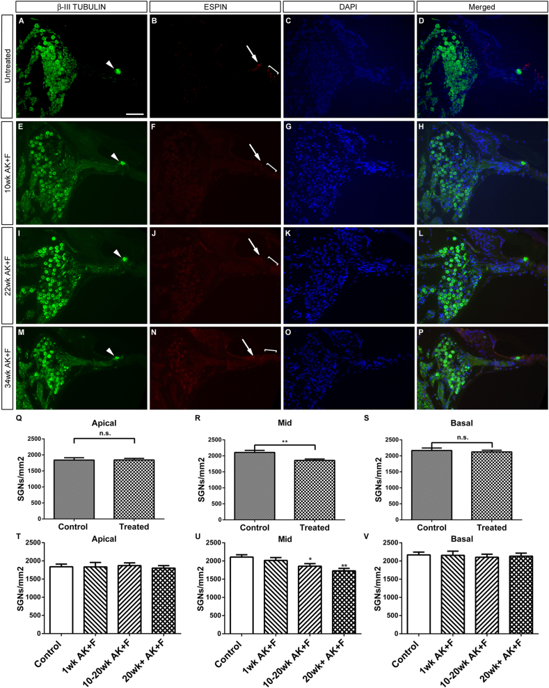
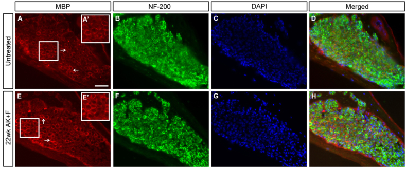
The Mongolian gerbil, Meriones unguiculatus, has been widely employed as a model for studies of the inner ear. In spite of its established use for auditory research, no robust protocols to induce ototoxic hair cell damage have been developed for this species. In this paper, we demonstrate the development of an aminoglycoside-induced model of hair cell loss, using kanamycin potentiated by the loop diuretic furosemide. Interestingly, we show that the gerbil is relatively insensitive to gentamicin compared to kanamycin, and that bumetanide is ineffective in potentiating the ototoxicity of the drug. We also examine the pathology of the spiral ganglion after chronic, long-term hair cell damage. Remarkably, there is little or no neuronal loss following the ototoxic insult, even at 8 months post-damage. This is similar to the situation often seen in the human, where functioning neurons can persist even decades after hair cell loss, contrasting with the rapid, secondary degeneration found in rats, mice and other small mammals. We propose that the combination of these factors makes the gerbil a good model for ototoxic damage by induced hair cell loss.


Figures

Fig. S1
Fig. S1
Phase contrast imaging demonstrates the loss of the hair cells in kanamycin/furosemide treated animals. Untreated (A–D, I–L, Q–T) and kanamycin/furosemide treated animals at one week (E–H; M–P; U–X) were stained for the presence of the calcium binding protein PARVALBUMIN, which marks the inner hair cells in intact animals (asterisks, B, J, R), and the sensory afferents (see Fig. S2 for further demonstration of this); whereas only the nerve fibres are labelled in the treated samples (arrowheads, F, N, V). Phase microscopy imaging demonstrates the loss of inner (asterisks, A, I Q) and outer hair cells (brackets, A, I Q). Representative apical (A–H), mid (M–P) and basal (Q–X) sections are shown, and are counterstained with DAPI (G, K, O, S, W). Scale bar – 50 μm throughout.
Fig. S2
Fig. S2
Sensory afferents do not degenerate in the absence of inner hair cells. Untreated (A–D, I–L, Q–T) and kanamycin/furosemide treated animals at different time points (1wk, E–H; 13wk M–P; 37wk U–X) were co-stained for the presence of the sensory afferent marker Na, K-ATPase α3 subunit (NKAα3) and the calcium-binding protein PARVALBUMIN (as per Fig. S1). The latter marks both the sensory afferents and the inner hair cells in intact animals (arrowheads and asterisks, B, J, R), whereas only the nerve fibres are labelled in the treated samples (arrowheads, F, N, V). Colabelling with the marker NKAα3 (arrowheads, A, E, I, M, Q, U) again demonstrates the presence of fibres within the damaged organ of Corti, even at 37wk post-treatment. Similar results were seen at apical (A–H), mid (Q–X) and basal (I–Q) levels of the cochlea. There is also innervation of the OHCs (square brackets, A, I, Q) by PARVALBUMIN positive fibres (arrows, B, J, R) – this staining is either lost (F, V) or degenerative (N) in treated animals, in which the outer hair cells have been lost, suggesting that the preservation of the sensory afferents might be unique. Scale bar – 50 μm throughout.
Fig. 1
Fig. 1
Topical application of gentamicin has no effect on the hearing organ. Gentamicin sulphate solution was applied directly to the round window membrane either acutely (E–H, Q) or as in a ‘slow release’ paradigm on a gelatine sponge (M–P, R). Immunofluorescence for β-III TUBULIN and the hair cell markers MYO7A (B, D, F, H) and ESPIN (J, L, N, P) show that there is no effect on the organ of Corti in either situation – comparison with untreated contralateral ears (A–D; I–L) shows no overt difference in structure or integrity of the inner and outer hair cells. Sections are counterstained with DAPI to delineate cell nuclei. There is no disruption to the innervation of the IHCs by spiral ganglion neurons (arrowheads, A, E, I, M). Click ABR measurements were taken at intervals post-treatment – no difference in response was found down to 20 dB with either treatment regime (Q, R). Arrows point to inner hair cells, brackets delineate the three rows of outer hair cells. Scale bar – 50 μm.
Fig. 2
Fig. 2
Application of kanamycin and bumetanide has minor effects on hearing. A combinatorial approach using kanamycin and bumetanide has no effect when dosed systemically (A–D, I), and only gives a moderate loss of hearing when given as a topical, chronic application (E–H, J–L). Immunofluorescence for ESPIN shows that in both cases the organ of Corti is intact (arrows and brackets B, F, compare with untreated control, Fig. 1J, L) and β-III TUBULIN staining shows that the innervation to the hair cells is normal (arrowheads, A, E). Sections are counterstained with DAPI to delineate cell nuclei. Typical sections from each sample are shown – apical turn (A–D), basal turn (E–H). Click ABR responses are maintained at 20 dB in the systemic treatment (I). There is a minor increase in auditory threshold in the topical case 6wk after treatment (J, K), and this functional decrement is most pronounced in the mid-frequency range, from 18 to 30 kHz (L). Arrows point to inner hair cells, brackets delineate the three rows of outer hair cells. Scale bar – 50 μm.
Fig. 3
Fig. 3
Systemic application of kanamycin and furosemide destroys inner and outer hair cells. Systemic application of kanamycin and furosemide gives a rapid and comprehensive destruction of both inner and outer hair cells. Immunofluorescence for the hair bundle marker ESPIN shows a complete loss of immunoreactivity 1wk after drug administration, implying a loss of hair cell function (compare arrows and brackets in B with the control situation in Fig. 1J). The tunnel of Corti is present at this stage (t, A, D). The hair bundle loss is permanent – at both 16wk and 34wk post-treatment, there is no restoration of the hair bundle (arrows and brackets, F, J) and the organ of Corti has degenerated – asterisks in F, H point to a small area of anomalous espin expression which would not constitute a functioning hair cell and may represent a hair cell in the process of being engulfed and destroyed. However, the presence of the spiral ganglion neuron processes remains intact (arrowheads, A, E, I.) DAPI is used as a counterstain as previously. Average click ABR thresholds rise considerably after treatment (M), from around 27 dB initially to around 97 dB after seven days. No difference is seen between the responses of the left and right ears (left ear n = 9; ****p < 0.0001; right ear n = 15; ****p < 0.0001). Pure tone ABR threshold measurements one week after kanamycin/furosemide treatment demonstrate an equivalent loss of response across the frequency range measured, 2–32 kHz (N) (n = 6; ****p < 0.0001). Mean and SEM plotted throughout. Scale bar – 50 μm.
Fig. 4
Fig. 4
Quantification of hair cell loss at all three cochlear levels. Whole-mount immunostaining of the cochlea with antibodies directed towards βIII-TUBULIN as a marker for spiral ganglion neurons (A, D, G, J, M, P and merged panels C, F, I, L, O, R) and MYO7A (B, E, H, K, N, Q and merged panels) to show the hair cells demonstrates a marked and quantifiable loss of both IHCs and OHCs at one week after kanamycin/furosemide treatment (D–F, J–L, P–R) when compared with untreated controls (A–C, G–I, M–O). Apical (A–F) mid (G–L) and basal (M–R) turns have an equivalent loss of OHCs, whereas there is a handful of IHCs remaining basally (arrows, Q) – however, these cells have lost their elongated morphology and are likely to represent a dying population. Cell counts were carried out and the mean number of each cell type per turn is displayed graphically in S, T; error bars represent SEM Scale bar – 25 μm throughout A–R.
Fig. 5
Fig. 5
Expression of the markers OCP2 and αTUBULIN in drug treated animals. The proteins OCP2 and acetylated αTUBULIN are markers for non-sensory supporting cells in the organ of Corti. Their expression remains high in the cochlea at all stages post-treatment with kanamycin/furosemide. OCP2 levels are comparable with the untreated condition (A–C) at 1wk (D–F), 22wk (G–I) and 34wk (J–L), suggesting that the damaged epithelium retains some level of differentiation, although it has become flattened in appearance by 34wk (J–L). Asterisks in A, C, D, F, G, I mark the intact tunnel of Corti; arrows in J, L indicate where this structure has collapsed. Costaining of MYO7A with αTUBULIN (M–X) shows a similar profile. In the untreated condition (M–P), the inner hair cells are clearly visible (arrowheads, N, P) – this staining is lost at all stages post-treatment as before (Q–X). The support cells, marked by αTUBULIN, remain intact and differentiated (arrows, M, P, Q, T, U, X) and the tunnel of Corti remains open (Q–X) up to 22wk post-treatment (asterisks, M, P, Q, T, U, X). DAPI is used as a nuclear counter-stain. Scale bar – 50 μm throughout.
Fig. 6
Fig. 6
SGNs are preserved well after long-term kanamycin/furosemide-induced ototoxicity. SGNs residing in Rosenthal's canal were stained for the marker β-III TUBULIN and counted in untreated (A, D) and treated animals (E, H, I, L, M, P) at various time points post drug treatment. In all treated cases, immunostaining with the marker ESPIN demonstrates a loss of both inner (arrows – F, J, N) and outer hair cells (brackets – F, J, N) compared to the untreated situation (B); however in all cases, β-III TUBULIN staining suggests that innervation to the degenerating organ of Corti is maintained (arrowheads, E, I, M) as before (Fig. 3). There is no significant difference in the remaining number of SGNs at apical and basal levels of the cochlea, with a significant but mild loss in the mid-turn. (Q–S; **p < 0.05). A breakdown of the SGN counts between different groups at the different time points (T–V) demonstrates that the extent of SGN loss in the mid-turn occurs at 10–20wk post-treatment, becoming slightly worse at 20wk+ (U). Mean and SEM are plotted. Representative images for mid-apical turns are shown for each condition, with DAPI used as a nuclear counterstain. Scale bar – 100 μm, panels A–P.
Fig. 7
Fig. 7
Sensory afferent neural processes remain into the organ of Corti in the absence of inner hair cells. Untreated (A–D, I–L, Q–T) and kanamycin/furosemide treated animals at different time points (1wk, E–H; 13wk M–P; 37wk U–X) were co-stained for the presence of the presynaptic ribbon component RIBEYE/CtBP3 and the sensory afferent marker Na, K-ATPase α3 subunit (NKAα3). In untreated samples, there is clear localisation of the RIBEYE protein at the base of the IHCs (arrows, A’, I’, Q’; apical, basal and mid turns respectively). This staining is mostly lost 1wk post-treatment (arrow, E’) and has completely gone at later stages (13wk, M’; 37wk, U’). There is a concomitant loss in treated samples of the nuclear localised RIBEYE protein which is seen in the IHCs of untreated samples (asterisks, A, A’; I, I’; M, M’), indicating a loss of these cells. The OHCs, indicated by brackets in A, D, I, L, Q, S, are also lost in the treated conditions. The SGN marker NKAα3 is maintained in all samples regardless of time post-treatment (arrowheads B, F, J, N, R, V), further confirming the residual ‘innervation’ of the organ of Corti months after the loss of the IHCs. Panels A’, E’, I’,M’, Q’ and U’ are magnified insets of the boxed regions in A, E, I, M, Q and U. Scale bars – 50 μm throughout panels A–X, while 10 μm in panels A’, E’, I’, M’, Q’, U’.
Fig. 8
Fig. 8
SGNs do not demyelinate in the long-term after kanamycin/furosemide treatment. Untreated (A–D) and a 22wk post-treatment (E–H) sample were examined for the expression of myelin basic protein (MBP, A, D, E, H) in Rosenthal's canal of the basal turn of the cochlea. Arrows (A, E) point to MBP staining ensheathing the SGNs. Inset boxes in A’, E’ represent 1.5× digitally magnified images of the corresponding boxed regions in A and E, respectively to show detail of the MBP sheath. There is no difference in expression apparent in the treated vs. the untreated condition. Counterstaining with neurofilament-200 (NF-200; B, F) showed the presence of nerve fibres in both cases; DAPI is used to counterstain the cell nuclei (C, D, G, H). Scale bar – 50 μm throughout.

Similar articles

Cited by

References

    1. Abrashkin K.A., Izumikawa M., Miyazawa T., Wang C.H., Crumling M.A., Swiderski D.L., Beyer L.A., Gong T.W., Raphael Y. The fate of outer hair cells after acoustic or ototoxic insults. Hear Res. 2006;218:20–29. - PubMed
    1. Alam S.A., Ikeda K., Kawase T., Kikuchi T., Katori Y., Watanabe K., Takasaka T. Acute effects of combined administration of kanamycin and furosemide on the stria vascularis studied by distortion product otoacoustic emission and transmission electron microscopy. Tohoku J. Exp. Med. 1998;186:79–86. - PubMed
    1. Aran J.M., Darrouzet J. Observation of click-evoked compound VIII nerve responses before, during, and over seven months after kanamycin treatment in the guinea pig. Acta Otolaryngol. 1975;79:24–32. - PubMed
    1. Astbury P.J., Read N.G. Kanamycin induced ototoxicity in the laboratory rat. A comparative morphological and audiometric study. Arch. Toxicol. 1982;50:267–278. - PubMed
    1. Bae W.Y., Kim L.S., Hur D.Y., Jeong S.W., Kim J.R. Secondary apoptosis of spiral ganglion cells induced by aminoglycoside: Fas-Fas ligand signaling pathway. Laryngoscope. 2008;118:1659–1668. - PubMed

Publication types